Journal List > Korean Circ J > v.41(11) > 1016786

Hachinohe, Jeong, Kim, Cho, Ahmed, Hwang, Lee, Sim, Park, Kim, Hong, Ahn, and Kang: Drug-Eluting Stent as an Option for Intractable In-Stent Coronary Restenosis

Abstract

A 51-year-old man was admitted due to an acute anterior ST-segment elevation myocardial infarction. After thrombolytic therapy using recombinant tissue plasminogen activator, stent implantation was performed from the proximal left anterior descending artery (LAD) to the mid LAD using a bare-metal stent (BMS). Since then, the patient suffered five repeated episodes of in-stent restenosis (ISR). At the first ISR, he was treated with plain old balloon angioplasty (POBA). At the second ISR, he was treated with brachytherapy, and at the third ISR, he was treated with POBA and one more BMS distal to the previously implanted stent. At the forth, only POBA was performed, and finally, at the fifth ISR, a sirolimus-eluting stent was implanted. Following that, the patient remained asymptomatic and follow-up coronary angiography showed no ISR.

Introduction

Stents reduce clinical and angiographic restenosis compared to conventional balloon angioplasty.1) However, in a number of patients with implanted stents, neointimal hyperplasia produces in-stent restenosis (ISR).2) Although often considered a benign process, recent data reveals that ISR has a negative impact on long-term survival.3) In fact, repeated procedures have been associated with a higher risk of myocardial infarction and stent thrombosis.4) Vascular brachytherapy was the approved therapy, in comparison with balloon angioplasty,5)6) but recent studies suggest that drug-eluting stents (DES) may be superior to brachytherapy.7)8) We experienced one intractable ISR case which repeated regardless of successful brachytherapy treatment, and which was treated using DES.

Case

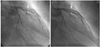
On February 2000, a 51-year-old man was admitted to the hospital with an acute anterior ST-segment elevation myocardial infarction. His electrocardiogram (ECG) showed ST-segment elevation in V1-5. After thrombolytic therapy using recombinant tissue plasminogen activator, stent implantation was performed from the proximal left anterior descending artery (LAD) to the mid LAD using 3.0×27 mm bare-metal stent (BMS) as an elective percutaneous coronary intervention (Fig. 1).
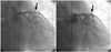
One year later, recurrent chest pain developed and an ECG showed ST-segment elevations in V1-5. Coronary angiography (CAG) revealed type IV (total) ISR in the proximal LAD, which was treated with balloon angioplasty using a 3.0×20 mm balloon (the first ISR). One year after that, recurrent chest pain developed, and ECG showed no ST-T changes, but CAG revealed type II (diffuse) ISR. This second ISR was treated with brachytherapy with a Rhenium filled balloon (300 seconds at 6 atm using a 3.0×20 mm balloon) (Fig. 2).
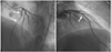
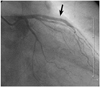
Four months later, recurrent chest pain developed once again and an ECG showed ST-segment elevations in V1-4. CAG revealed type IV ISR secondary to brachytherapy failure, which was treated with 3.0×20 mm balloon and 3.0×18 mm BMS implantation distal to the previously implanted BMS (the third ISR) (Fig. 3). Three months later, chest pain recurred and ECG showed ST-segment elevations in V3-5. CAG revealed type II ISR, which was treated with only ballooning (the fourth ISR). Nine months later, recurrent chest pain developed, and ECG showed no ST-T changes. CAG revealed type II ISR in the proximal and mid LAD stent. A 3.0×33 mm Cypher stent® (Cordis Johnson & Johnson, Bridgewater, NJ, USA) was implanted to treat this fifth ISR presentation (Fig. 4). The patient then remained asymptomatic and the 5-year follow-up CAG showed no ISR (Fig. 5).

Discussion

Bare-mental stent has improved procedural success and long-term outcomes, and reduced clinical and angiographic restenosis compared to plain old balloon angioplasty (POBA).1)9) Although stenting can resolve elastic recoil, negative remodeling and dissection caused by POBA and neointimal hyperplasia produces ISR in 20% to 50% of the patients implanted stent.2)10) ISR is often considered to be a benign process, but a recent report showed that it presents with acute myocardial infarction or unstable angina in over one-third of cases3) and has a negative impact on long-term survival.11) In fact, repeated procedures have been associated with a higher risk of myocardial infarction and stent thrombosis.4)
There are many treatment options for ISR. Although POBA is the first-line option, its results have been disappointing. Repeated use of BMS also exacerbates the risk of recurrence. Coronary artery bypass surgery may be an option for the treatment of recurrent ISR after BMS implantation. Alternative options, such as rotational atherectomy, excimer laser angioplasty, and cutting balloon did not reduce recurrent ISR.12)
Several reports show the efficacy of brachytherapy in patients with ISR,5)6)13)14) but brachytherapy presents problems such as logistical complexity, expense, and durability.12) Recent studies suggest that DES is superior to POBA,8)15) and moreover DES may even be superior to brachytherapy.7)8) This is a unique case for the successful management of intractable ISR with DES, even after POBA, BMS and brachytherapy.
In conclusion, we experienced an intractable ISR case, which can be treated by using DES. As recent studies show, DES is the best treatment option for ISR, and may even be used successfully to treat brachytherapy failure and intractable ISR.

Figures and Tables

Fig. 1
A diagnostic coronary angiogram (CAG) revealed successful thrombolysis status but a critical stenosis of the proximal and mid left descending artery. After successful stent implantation using a 3.0×27 mm bare-metal stent, final CAG showed no residual stenosis with good distal flow.
kcj-41-677-g001
Fig. 2
Second in-stent restenosis (ISR). A coronary angiogram (CAG) revealed type II ISR. Brachytherapy with Rhenium was performed for 300 seconds at 6 atm using a 3.0×20 mm balloon. Final CAG showed no residual stenosis with good distal flow.
kcj-41-677-g002
Fig. 3
Third in-stent restenosis (ISR). A coronary angiogram (CAG) revealed type IV ISR secondary to brachytherapy failure. Plain old balloon angioplasty using a 3.0×20 mm balloon and 3.0×18 mm bare-metal stent implantation distal to previously implanted stent was performed. Final CAG showed no residual stenosis with good distal flow.
kcj-41-677-g003
Fig. 4
Fifth in-stent restenosis (ISR). A coronary angiogram (CAG) revealed type II ISR in proximal and mid left anterior descending artery stent. A 3.0×33 mm Cypher stent® (Cordis Johnson & Johnson) was inplanted. Final CAG showed no residual stenosis with good distal flow.
kcj-41-677-g004
Fig. 5
On follow-up coronary angiogram performed one week after stenting, the stent in the left anterior descending artery was patent without narrowing of the ostium of left circumflex and distal left main artery.
kcj-41-677-g005

Notes

The authors have no financial conflicts of interest.

References

1. Serruys PW, de Jaegere P, Kiemeneij F, et al. Benestent Study Group. A comparison of balloon-expandable-stent implantation with balloon angioplasty in patients with coronary artery disease. N Engl J Med. 1994. 331:489–495.
2. Ahmed JM, Mintz GS, Weissman NJ, et al. Mechanism of lumen enlargement during intracoronary stent implantation: an intravascular ultrasound study. Circulation. 2000. 102:7–10.
3. Chen MS, John JM, Chew DP, Lee DS, Ellis SG, Bhatt DL. Bare metal stent restenosis is not a benign clinical entity. Am Heart J. 2006. 151:1260–1264.
4. Stone GW, Ellis SG, Colombo A, et al. Offsetting impact of thrombosis and restenosis on the occurrence of death and myocardial infarction after paclitaxel-eluting and bare metal stent implantation. Circulation. 2007. 115:2842–2847.
5. Waksman R, Bhargava B, White L, et al. Intracoronary beta-radiation therapy inhibits recurrence of in-stent restenosis. Circulation. 2000. 101:1895–1898.
6. Popma JJ, Suntharalingam M, Lansky AJ, et al. Randomized trial of 90Sr/90Y beta-radiation versus placebo control for treatment of in-stent restenosis. Circulation. 2002. 106:1090–1096.
7. Stone GW, Ellis SG, O'Shaughnessy CD, et al. Paclitaxel-eluting stents vs vascular brachytherapy for in-stent restenosis within bare-metal stents: the TAXUS V ISR randomized trial. JAMA. 2006. 295:1253–1263.
8. Alfonso F, Pérez-Vizcayno MJ, Hernandez R, et al. A randomized comparison of sirolimus-eluting stent with balloon angioplasty in patients with in-stent restenosis: results of the restenosis intrastent: balloon angioplasty versus elective sirolimus-eluting stenting (RIBS-II) trial. J Am Coll Cardiol. 2006. 47:2152–2160.
9. Fischman DL, Leon MB, Baim DS, et al. Stent Restenosis Study Investigators. A randomized comparison of coronary-stent placement and balloon angioplasty in the treatment of coronary artery disease. N Engl J Med. 1994. 331:496–501.
10. Kastrati A, Mehilli J, Dirschinger J, et al. Restenosis after coronary placement of various stent types. Am J Cardiol. 2001. 87:34–39.
11. Schühlen H, Kastrati A, Mehilli J, et al. Restenosis detected by routine angiographic follow-up and late mortality after coronary stent placement. Am Heart J. 2004. 147:317–322.
12. Radke PW, Kaiser A, Frost C, Sigwart U. Outcome after treatment of coronary in-stent restenosis; results from a systematic review using meta-analysis techniques. Eur Heart J. 2003. 24:266–273.
13. Hong YJ, Jeong MH. New methods of vascular brachytherapy for coronary stent restenosis. Korean Circ J. 2003. 33:967–976.
14. Kim SH, Kim MH, Cha KS, et al. Effect of vascular brachytherapy using HolmiuM-166 liquid balloon system after cutting balloon angioplasty in patients with stent restenosis (BRAHMS Study). Korean Circ J. 2005. 35:591–596.
15. Kastrati A, Mehilli J, von Beckerath N, et al. Sirolimus-eluting stent or paclitaxel-eluting stent vs balloon angioplasty for prevention of recurrences in patients with coronary in-stent restenosis: a randomized controlled trial. JAMA. 2005. 293:165–171.
TOOLS
Similar articles