

 PDF
PDF ePub
ePub Citation
 Print
Print


Introduction
Slow coronary flow (SCF) is characterized by delayed contrast dye opacification without significant stenosis of epicardial coronary arteries. Most patients with SCF are debilitated with recurrent chest pain, commonly resulting in repeated hospital admissions.1) However, the pathophysiology and clinical implications of SCF are not fully understood. It is likely that endothelial inflammation and microvascular vasomotor dysfunction are associated with SCF pathophysiology.2-8) Some reports have suggested that SCF might be caused by atherosclerotic changes in the coronary artery microvasculature and could be an early marker of subclinical atherosclerosis.9)
Non-invasive and simple diagnostic tools have been developed to detect subclinical atherosclerosis. Intima-media thickness (IMT) of the carotid artery reflects atherosclerotic changes and strongly correlates with the presence and extent of coronary artery disease.10)11) Arterial stiffness is also an important aspect of atherosclerosis related to vessel function that can be evaluated noninvasively by measuring pulse wave velocity (PWV), which is reportedly associated with the severity of vascular damage.12) Many studies in different populations have shown that these are good parameters that indicate the degree of vascular atherosclerosis.11)12) We hypothesized that SCF is related to coronary artery atherosclerosis, so we compared carotid IMT and PWV between an SCF and normal coronary flow (NCF) patient groups.
Subjects and Methods
Study population
The study was conducted from August 2005 to September 2006. We included 101 consecutive patients who complained of chest pain but showed no significant coronary stenosis on coronary angiography. A complete history, as well as physical and laboratory examinations, were obtained from all patients before performing the coronary angiography. Smokers were defined as subjects who had smoked regularly for more than 1 year. Hypertension was defined as systolic blood pressure ≥140 mm Hg or diastolic blood pressure ≥90 mm Hg or current use of antihypertensive agents. Diabetes was defined as the use of insulin, oral hypoglycemic agents, or a fasting plasma glucose level of ≥126 mg/dL. After the coronary angiogram, we confirmed the clinical diagnosis and categorized the patient as follows: acute myocardial infarction, unstable angina, microvascular angina, noncardiac chest pain, or congestive heart failure. The definition of acute myocardial infarct was a typical rise and/or gradual fall (troponin) or a more rapid rise and fall (creatine kinase-MB fraction) of biochemical markers of myocardial necrosis, with at least one ischemic symptom (chest pain) or electrocardiogram (ECG) changes indicative of ischemia. Unstable angina was considered as ischemic symptoms (chest pain) and no elevation in bio-chemical markers with ECG changes (new 1 mm or greater ST-segment depression or T-wave inversion in multiple precordial leads). The definition of microvascular angina required ischemic symptoms (chest pain) but no elevation in biochemical markers with the following ECG changes: ST-segment depression of 0.5-1 mm, T-wave flattening, or inver-sion <1 mm in the lead with a dominant R wave. The non-cardiac chest pain group complained of ischemic symptoms (chest pain) but no elevation in biochemical markers and a normal or unchanged ECG. Congestive heart failure was diagnosed clinically using the Framingham criteria.
Coronary angiography and thrombolysis in myocardial infarction frame count
Coronary angiography was performed with the radial or femoral approach using the standard Judkins technique. Coronary arteries were shown in the left and right oblique planes and in cranial and caudal angulations. All angiograms were filmed at 15 frames per second. The Thrombolysis in Myocardial Infarction frame count (TIMI frame count, TFC) was obtained by the method of Gibson et al.13) The first frame used for counting was the first frame in which dye fully entered the artery. The last frame counted was the one in which dye first entered the end point branch off the target artery. The follow-ing distal landmark branches of the target artery were used for the analysis: the distal bifurcation in the left anterior descending artery (LAD), the distal bifurcation of the segment with the longest total distance in the left circumflex artery (LCX), and the first branch of the posterolateral artery in the right coronary artery (RCA). Because the length of the LAD is anatomically longer than that of the LCX and RCA, the LAD frame counts were corrected by dividing by 1.7 to obtain a corrected TFC, as previously reported.13) We calculated the mean TFC for the LAD, LCX, and RCA. All participants with a mean TFC greater than two standard deviations from mean TFC in the noncardiac chest pain subgroup were considered to have SCF, whereas those with a value below this threshold were considered to have NCF. The threshold TFC value was 25 frames.
Intimamedia thickness and pulse wave velocity
The carotid IMT was calculated by ultrasound equipment with a linear transducer (12-MHz, Vivid 7 dimension, GE Healthcare, Piscataway, NJ, USA). The right and left common carotid arteries were scanned in a longitudinal plane with loss of the parallel configuration of the near and far walls of the common carotid artery. The reference point for measuring IMT was the beginning of the dilatation of the carotid bulb in the bifurcation area. The IMT was measured manually as the thickest point of the transition zone between the lumenintima and media-adventitia, 1 cm proximal to the reference point along at least 1 cm of axial length.14) The values for each carotid artery were averaged to obtain the mean carotid IMT. Carotid IMT was assessed by two cardiologists blinded to other clinical data. Disagreements were resolved by a third observer.
We measured PWV on the left and right side using a Vasera VS-1000 instrument (Fukuda Denshi Co. Ltd, Tokyo, Japan), which simultaneously records PWV, blood pressure, ECG and heart sounds. The PWV is theoretically calculated as the distance from two sites over the pulse transit time. The time interval between the brachium and the ankle (ΔTba; pulse wave transit time) was defined as the time interval between the wave front of the brachial waveform and that of the ankle wave form. The distance between sampling points was calculated automatically according to subject height. The path length from the heart to the brachium (Lb) was expressed using the following equation: Lb=0.2195×height of the patient (cm)-2.0734.15) The path length from the heart to the ankle (La) was expressed using the following equation: La=0.8129×height of the patient (cm)+12.328.15) Finally, the PWV was calculated from the following equation: PWV=(La-Lb)/ΔTba.15) The mean PWV value was calculated as the average of both PWV values.
Statistics
The statistical analysis was performed using the general linear model in JMP7.0 software (SAS Inc., Cary, NC, USA). The categorical variables were expressed as counts and percents. The categorical variables were compared with Pearson's chi-square test and an analysis of variance. Continuous variables are expressed as the mean±standard deviation (SD), and the independent t-test was used to compare the two groups. The Pearson's correlation test was used to determine the correlation between the TFC and the carotid IMT.
Results
Baseline characteristics
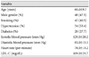
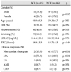
The clinical characteristics of the 101 patients in this study are summarized in Table 1. Overall, 54% of the subjects had hypertension, 28% had diabetes mellitus (DM), and 42% were smokers. Eighty-six patients were categorized into the NCF group, and 15 patients in the SCF group. Male patients (n= 11, 73.3%) were significantly more common in the SCF group than those in the NCF group (n=37, 43.0%, p<0.05). However, age, presence of DM, hypertension, and lowdensity lipoprotein levels did not differ between the NCF and SCF groups. Smoking tended to be more common in the SCF group, but the difference was not statistically significant (Table 2).
Clinical diagnosis and thrombolysis in myocardial infarction frame count
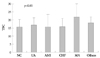
A significant difference was observed in the clinical diagnoses between the SCF and NCF groups. Microvascular angina was the most common clinical diagnosis (n=11, 73.3%) in the SCF group, whereas non-cardiac chest pain was the most common diagnosis in the NCF group (n=41, 47.7%). The TFC was 28.8±3.5 frames in the SCF group and 15.7±4.5 frames in the NCF group (Table 2). In particular, the TFC increased significantly in a subgroup with microvascular angina (21.8± 7.8 frame count), compared to that in the other clinical subgroups (Fig. 1). We also compared the TFC according to risk factors. The TFC in male patients tended to be higher than that of female patients (18.9±7.0 vs. 16.6±5.5, respectively), but the result was not statistically significant. The TFC was not different with the presence of other risk factors, such as DM, hypertension, or smoking.
Carotid intima-media thickness and pulse wave velocity
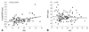
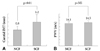
Carotid IMT was positively correlated with TFC (r=0.40, p<0.001), but no significant correlation was found between PWV and TFC (Fig. 2). Carotid IMT increased significantly in the SCF group compared to that in the NCF group (1.2± 0.3 mm and 0.8±0.1 mm, respectively, p<0.01). However, no difference was found in the PWV between the SCF and NCF groups (SCF; 14.1±2.4 m/s, NCF; 14.5±2.8 m/s) (Fig. 3).
Discussion
After the first description of the SCF phenomenon in six patients with typical angina pectoris by Tambe et al.2) SCF has been widely recognized during routine coronary angiography. SCF reveals various clinical presentations such as unstable angina, acute myocardial infarction, ventricular tachycardia, hypertrophic cardiomyopathy, and microvascular angina.1)16)17)
Camsari et al.9) used intravascular ultrasound to show that diffuse intimal thickening of the coronary arteries and carotid IMT are correlated with TFC in patients with SCF.18) Some reports have suggested that homocysteine, a well-known factor associated with generalized atherosclerosis, increases in patients with SCF.14) In other reports, simvastatin restores endothelial function and improves myocardial perfusion in patients with SCF.19) These findings suggest that diffuse atherosclerosis may play a role in the pathophysiology of SCF. Similar to previous studies, our findings showed that TFC was positively correlated with the degree of carotid IMT, and that carotid IMT increased more significantly in the SCF group than that in the NCF group. According to these findings, it seems that the degree of atherosclerosis is a determining factor of SCF.
In our study, SCF was defined as a TFC greater than 25 frames (filmed at 15 frames per second). This is extreme SCF compared to previous studies.9)13)14) IMT of the carotid arteries in the SCF group increased greater in our results than those of other reports.13)14)20) These findings indicate that our subjects were more severe cases of SCF.
The results also showed no difference in the PWV between the SCF and NCF groups. PWV is considered a good indicator of the degree of atherosclerosis; however; compared to IMT, it seems to be less sensitive for coronary artery atherosclerosis. Similar to the present study, research on the relationship between carotid IMT, brachial-ankle PWV (baPWV), and the severity of coronary artery atherosclerosis, showed a relatively good correlation between carotid IMT and the Gensini score, which is a measure of the coronary artery disease severity, whereas baPWV correlated only weakly with this score.21) That is, carotid IMT reveals the degree of atherosclerosis in coronary arteries, and it may be more suitable for evaluating coronary microcirculation than baPWV.21-23) However, the reason for the different correlation in carotid IMT and baPWV is unclear. Therefore, further study is needed for confirmation.
Because SCF is associated with coronary atherosclerosis, it can be influenced by various cardiovascular risk factors such as gender, age, DM, hypertension, hyperlipidemia, and smoking. However, no significant differences were observed for age, gender, hypertension, DM, smoking, and total cholesterol in a series of 50 patients with SCF.9) In another report, 47 patients with SCF also showed no significant differences in risk factors from the control group.1) Furthermore, we found no significant difference in the risk factors between the SCF and the NCF groups, except for gender. Although conventional cardiovascular risk factors reflect an advanced stage of atherosclerosis, we found no significant difference in conventional cardiovascular risk factors between the SCF and NCF groups. Thus, it is possible that risk factors other than conventional cardiovascular risk factors are present in patients with SCF. This should be explored further in a larger study.
There were some limitations to our study. First, we showed a correlation between SCF and carotid IMT and the possibility of atherosclerosis in coronary microvasculature, but we did not directly assay the myocardial microvasculature itself to evaluate the degree of atherosclerosis. A pathological comparison in patients with SCF will help in the understanding of the SCF mechanism. Second, because we included a small number of patients, some difficulties were encountered when identifying the characteristics of the cardiovascular risk factors. A large multi-center based clinical trial is needed to identify clinical characteristics and to manage patients with coronary microvascular dysfunction.
 XML Download