

 PDF
PDF ePub
ePub Citation
 Print
Print


Introduction
Aortic stenosis (AS) is a gradually progressive disease that is characterized by an increase in calcium deposition and this leads to progressive narrowing of the aortic valve (AV). AS is mediated by a chronic inflammatory disease process that is very similar to that seen in atherosclerosis,1-4) but lipid-lowering therapy did not slow the progression of AS in the SALTIRE, SEAS or ASTRONOMER trials.5-7) However, the cited trials may have targeted patients in whom the disease was too advanced for lipid-lowering therapy to be effective,8) or in whom atherogenesis was not the central pathogenic process in AS.9) Even in a study that included patients with mild and moderate AS,10) hypercholesterolemia was not associated with adverse events, whereas the extent of AV calcification and the peak aortic jet velocity were associated with poor outcomes. Because the progression of AS is variable and this is influenced by the valve morphology 11) and the degree of stenosis,12) a prospective study is needed that will evaluate the natural history and predictors of progression in patients with mild AS. Using our prospectively collected registry data on patients with AS,11) we evaluated the long-term outcomes and predictors of echocardiographic progression in patients with mild AS.
Subjects and Methods
Study population
A prospective registry was created in our hospital, using a standard case report form, and this included all the consecutive patients with AS and who were undergoing echocardiography. The case report forms, including information on the patient demographics, the clinical presentation and the echocardiographic data, are stored in an electronic database.11) The clinical and echocardiographic follow-up data on the patients has been collected annually. From 2001 to 2003, a total of 103 consecutive asymptomatic patients (62.1±11.9 years; 31 males) with mild AS were enrolled. Mild AS was defined as AV thickening accompanied by a peak aortic jet velocity ≥2.0 and <3.0 m/sec. In agreement with the exclusion criteria of the SEAS trial,6) patients were excluded if they had other significant valvular disease, moderate or severe aortic regurgitation, a left ventricle (LV) ejection fraction (EF) <50%, regional wall motion abnormalities, established coronary, cerebral and/or peripheral vascular disease, renal insufficiency or uncontrolled endocrine, metabolic or liver disease.
Informed consent was obtained from each patient and the study protocol was approved by the Ethics Committees of our institutions.
Echocardiographic evaluation
All the patients underwent annual comprehensive two-dimensional and Doppler echocardiographic follow-up examinations using Hewlett-Packard Sonos 2500 or 5500 imaging systems equipped with 2.5-MHz transducers (Hewlett-Packard, Andover, MA, USA). The end-systolic dimension, end-diastolic dimension and end-diastolic interventricular septal and po-sterior wall thickness of the LV were measured from the pa-rasternal M-mode acquisitions. The end-systolic volume, end-diastolic volume and EF of the LV were obtained using the biplane Simpson method,13) and the LV mass was calculated with the formula validated by Devereux et al.14) On the two-dimensional imaging of the AV in the parasternal short axis view, the etiology of AS was defined as bicuspid if two leaflets were clearly visible during systole, as rheumatic if commissural fusion and mitral valve involvement were observed and as degenerative if thickening and calcification of the leaflets were notable.15) AV calcification was graded as mild when a single spot or scattered small spots of echo brightness were observed, as moderate when the calcified mass involved one or two leaflets and as severe when there was extensive thickening and the calcified mass involved all the leaflets.16) The maximal aortic jet velocity (AV Vmax) was recorded using the window (apical, right parasternal, or suprasternal) that yielded the highest velocity signal. The maximal and mean pressure gradients across the AV were calculated using a modified Bernoulli equation, and the AV area was estimated from the continuity equation using the LV outflow tract diameter and the flow velocity.15) For patients in sinus rhythm, the data from 3 cardiac cycles were averaged. However, for patients with irregular rhythm such as atrial fibrillation, 5 cycles were averaged to ensure the accuracy of the results. The change of the AV Vmax was calculated during annual echocardiographic follow-up, and the average change of the AV Vmax was determined for each patient. A rapid progression of AS was defined as an average annual increase of the AV Vmax ≥0.20 m/sec/year because the mean increase of the peak aortic jet velocity was 0.14 m/sec/year in the SEAS trial, which included 1,873 patients with mild-to-moderate AS.
Follow-up
The follow-up was completed and the data was collected until December 2009 during each patient's annual visit to the echocardiographic laboratory and by a detailed review of all the medical records and telephone interviews. Deaths were classified as cardiac or non-cardiac, based on the medical records. The endpoint of the study was defined as cardiac death or AV replacement (AVR) during follow-up. The progression of AS was monitored by the change of the AV Vmax on the annual echocardiographic follow-up.
Statistical methods
The continuous variables were compared using Student's t-test or the Wilcoxon rank sum test, and categorical variables were compared using the Chi-squared test or Fisher's exact test, as appropriate. Cumulative event curves were generated using the Kaplan-Meier method. Linear regression analysis was used to investigate the relationship between the continuous variables. Stepwise multiple regression analysis was employed to identify the independent predictors of AS progression. A univariate probability value threshold of 0.10 was set for entry into the equation. All the p-values were two-sided, and a p<0.05 was considered significant. All the statistical analyses were performed using Statistical Package for the Social Sciences (SPSS) version 12.0 for Windows (SPSS Inc., Chicago, IL, USA).
Results
Baseline characteristics
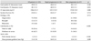
The etiology of AS was degenerative in 73 (70.9%) patients, rheumatic in 15 (14.6%) and bicuspid in 12 (11.7%). Fifty-six patients (54.4%) had hypertension, 4 (3.9%) had diabetes and 7 (6.8%) were current smokers. Forty-five patients (43.7%) had serum cholesterol concentrations >200 mg/dL and 44 (42.7%) had moderate-to-severe calcification of the AV.
Clinical and echocardiographic follow-up
During a median clinical follow-up time of 7.3 years (interquartile range: 6.6-8.4 years), there were two non-cardiac deaths and two cardiac events (both AVRs). The estimated 7-year cardiac event-free survival rate was 98±1%. One patient underwent AVR because of developing severe symptomatic AS, and one patient who underwent coronary artery bypass grafting combined with AVR because of the presence of severe AS. The causes of the two non-cardiac deaths were respiratory failure and suicide.
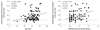
During a median echocardiographic follow-up time of 6.0 years (IQR: 4.4-6.5 years), the mean AV Vmax increased from 2.3±0.3 m/sec to 2.7±0.7 m/sec with an average annual increase of 0.08±0.10 m/sec/year. The annual increase in the AV Vmax was significantly related to the initial AV Vmax (r= 0.379, p<0.001) and age (r=0.252, p=0.011) (Fig. 1). On the stepwise multiple regression analysis, age, the baseline AV Vmax and moderate-to-severe calcification were the independent predictors of the annual progression rate (r=0.570, p< 0.001).
Relationship between the patient outcomes and the rate of progression
Rapid progression, defined as a ≥0.2 m/sec per year increase in the AV Vmax, was observed in 16 of 103 patients (15.5%) and slow progression (<0.2 m/sec per year) was observed in 87 patients (84.5%). The clinical and echocardiographic characteristics of these two groups are compared in Table 1 and 2. There were no significant between-group differences in age, gender, a history of diabetes or hypertension, the level of serum cholesterol, the LV dimensions, the LV mass index, the LV EF or the etiology of AS. However, the baseline AV Vmax (2.6±0.3 m/sec vs. 2.2±0.3 m/sec, respectively, p<0.001) and the incidence of moderate-to-severe AV calcification (92.9% vs. 36.5%, p<0.001) were significantly higher in the rapid progression group than in the slow progression group. Eight patients (50%) in the rapid progression group developed severe AS (AV Vmax ≥4.0 m/sec or AV area ≤0.75 cm2) during follow-up, and the 7-year cardiac event-free survival rate was lower in the rapid progression group than in the slow progression group (87.5±8.3% vs. 100%, respectively).
Discussion
We found that asymptomatic patients with mild valvular AS and who were prospectively followed-up by repeated echocardiography generally had a benign long-term prognosis and slow progression of AS. However, there was marked individual variability, and progression to severe AS was observed in 50% of the patients with rapid progression of AS.
The outcomes of the patients with mild and moderate AS have been reported to be worse than commonly assumed, indicating a need to consider rapid progression as predictive of excess mortality. The present study, which included only patients with mild AS and it excluded those with coronary, cerebral and/or peripheral vascular disease, showed that patients had a more benign natural history than was seen in a previous study.10) The cited works, which included patients with moderate-to-severe AS, found that the average rate of increase in the mean pressure gradient was about 8 mm Hg per year, the average decrease in the AV area was about 0.1 cm2 per year and the average rate of increase in the aortic jet maximum velocity ranged from 0.2 to 0.4 m/sec per year.5)17-20) We found that the average annual increase in the AV Vmax (0.08±0.10 m/sec/year) was lower than the 0.14±0.14 m/sec/year observed in the SEAS trial, which included patients with mild and moderate AS. This finding was similar compared with the recent data of mild AS in Korean patients.21) Our results suggest that more conservative approaches are needed for patients with mild AS.
We also found that the patient's age, the baseline AV jet velocity and the presence of moderate-to-severe calcification were independent predictors of AS progression. Previous studies have also reported that significant AV calcification, the peak aortic jet velocity and the rapid progression of AS were independent predictors of the outcomes for patients with mild and moderate AS. Our finding, that AV calcification was associated with a poor outcome, confirms the results of a study that utilized electron beam tomography to assess the degree of AV calcification.22) Our results suggest that echocardiographic observation alone is sufficient for patients with mild AS, but rapid progression is a risk factor for progressing to severe AS. Follow-up echocardiographic examinations of older patients and those with moderate-to-severe calcification or a higher baseline aortic jet velocity should be performed to detect rapid progression before severe symptoms occur.
Although AS was previously regarded as a degenerative change, this process is now recognized as sharing many histologic features with atherosclerosis and to be associated with the same risk factors.3)23)24) Thus, degenerative AS is currently regarded as an active disease similar to atherosclerosis, and it is progressive. Following the finding that hypercholesterolemia may be a risk factor for AS,25) several studies have assessed the effects of lipid-lowering therapy for halting or delaying progression of the disease.5)6)17-20) However, it is not clear if hypercholesterolemia affects the progression of mild AS, and we found that the serum cholesterol concentration was not associated with the annual increase of the aortic jet velocity even in patients with mild AS.
Limitations
The study patients were enrolled at a tertiary referral hospitals and the gender ratio, the incidence of hypertension and the LVMI might be different from other cohort studies. The number of study patients was not large enough to detect weak correlation between the cholesterol levels and progression rates, and to perform subgroup analysis with statistical testing of interactions according to the etiology of AS. The AV area was not measured in all the study patients because the progression of AS was monitored by the changes in the AV Vmax on the follow-up echocardiographic examinations. In addition, the predictors of the clinical outcomes could not be evaluated due to the very low rate of cardiac events in the patients with mild AS.
Conclusion
We have shown here that the progression of AS was slower than expected, and it was related to age, the baseline aortic jet velocity and AV calcification, but not with the serum cholesterol concentration in patients with mild AS. Because of the marked individual variability in the progression of AS, the patients showing rapid progression need close, regular follow-up.

 XML Download