

 PDF
PDF ePub
ePub Citation
 Print
Print


Introduction
The renin-angiotensin system (RAS) is one of the most important mechanisms implicated in the development of cardiovascular disease. The pathophysiological effects of angiotensin II, the key player in the RAS, that potentially lead to cardiovascular disease include vasoconstriction, endothelial dysfunction, increased thrombosis, superoxide production, vascular smooth muscle growth, myocyte hypertrophy, fibrosis, remodeling of tissues, and stimulation of a number of inflammatory and hormonal mediators.1)
Angiotensin-receptor blockers (ARBs) are the most recently approved class of antihypertensive agents that inhibit the RAS by selectively blocking the AT-1 receptor, so ARBs inhibit most of the deleterious effects of angiotensin II. In addition to blood pressure (BP) control, ARBs may have beneficial effects on left ventricular hypertrophy, arterial stiffness, vascular endothelial function, and carotid intima-media thickness (IMT), all of which are well-known surrogate markers and intermediate endpoints for cardiovascular morbidity and mortality.2)3)
Further, RAS blockers, including angiotensin-converting enzyme inhibitors (ACEIs) and ARBs, reduce the incidence of new-onset diabetes.4)5) Recent in vitro and animal studies have suggested that some ARBs act as partial peroxisome proliferator-activated receptor-gamma (PPARγ) agonists and that this does not depend on an AT-1 receptor blockade; thus, this may improve insulin sensitivity and the lipid profile.6)7)
Inflammation is one of the plausible mechanisms by which angiotensin II accelerates the development of atherosclerosis8) and evidence exists that inflammatory markers are reduced by ARBs independent of changes in BP.9)10)
The cardiovascular, metabolic, and anti-inflammatory effects of ARBs are thought to be similar and rarely compared within the drug class, though important structural differences exist among the various compounds. The purpose of this study was to compare these cardiovascular, metabolic, and anti-inflammatory effects between telmisartan and valsartan in uncomplicated hypertensive subjects.
Subjects and Methods
Subjects
We performed a prospective, randomized, open-label, fixed-monotherapy, single center, parallel-group study. Patients were recruited from the Cardiovascular Center at Korea University Guro Hospital, Seoul, Korea. Inclusion criteria were newly diagnosed or known hypertensive subjects who were not taking antihypertensive agents for more than the last 4 weeks. Patients with the following conditions were excluded; 1) hyperkalemia >5.5 mmol/L, 2) serum creatinine >2 mg/dL, 3) hemoglobin A1c (HbA1c) >8.0%, 4) secondary hypertension, 5) hormone therapy or steroid administration, 6) taking antidiabetic or lipid-lowering agents, 7) known coronary artery disease or significant atherosclerotic disease of other vessels and, 8) clinically significant nervous, gastrointestinal disease, or malignant disease. All subjects gave their written informed consent, and the Korea University institutional review board approved the study protocol in accordance with the Declaration of Helsinki of the World Medical Association.
After a 2-week run-in period, patients were randomized to receive either telmisartan (80 mg/day; n=30) or valsartan (160 mg/day; n=30) for a 12-week open-labeled phase. Patients were requested to take their study medication with water in the morning and at approximately the same time each day. Patients were seen on the screening visit, 1 week before randomization (baseline laboratory and cardiovascular studies), at entry (randomization), and at 4 and 12 weeks of treatment. At week 12, physical examinations, laboratory assessments, and cardiovascular studies were repeated.
Blood pressure measurement
After at least 10 minutes of rest, sitting systolic and diastolic BPs were recorded twice every 2 minutes by a research nurse using a random zero mercury sphygmomanometer fitted with an appropriate cuff size. The recorded BPs were averaged for use in the analysis. Patients in which systolic pressure was >140 mm Hg or diastolic pressure was >90 mm Hg were defined as having hypertension.
Anthropometric and laboratory measurements
Age, gender, body weight, waist circumference, and body mass index (BMI) were recorded. Waist circumference was measured at the midpoint between the lower border of the rib edge and the iliac crest, and BMI was calculated as the weight/height2 (kg/m2). Current smoking was defined as active smoking within the past 12 months. All blood samples were obtained in the morning with subjects in a fasting state. Insulin resistance was calculated by the Homeostasis Model of Assessment (HOMA).11) HbA1c was measured using high performance liquid chromatography (Bio-Rad Variant II, Hercules, CA, USA). Serum adiponectin and interleukin-6 levels were measured using an enzyme-linked immunosorbent assay (Samkwang Medical Laboratories, Seoul, Korea).
Measurement of pulse wave velocity
All procedures were conducted in a controlled environment by an experienced technician who was blinded to the clinical information. The carotid-femoral pulse wave velocity (cfPWV) was determined using the PP-1000 semiautomatic device (Hanbyul Meditech, Seoul, Korea).12) Briefly, the left common carotid artery (CCA), radial artery, femoral artery, and dorsal artery pressure waveforms were recorded non-in-vasively using a tonometric pressure sensor. An electrocardiogram, phonocardiogram and four-channel pressure wave-forms were simultaneously measured. The distance (D) tra-veled by the pulse wave was automatically obtained from the age- and height-based distance of the upper and lower extre-mity arteries of Koreans, and this was provided by the Korea Research Institute of Standards and Science. The pulse transit time (t), which was measured between the feet of the pressure waveforms that were recorded at two different recording points (the foot-to-foot method), was automatically determined. The PWV was automatically calculated as D/t. Am-ong the carotid-femoral, carotid-radial, and femoral-dorsal PWVs, we only used the cfPWV in the current study because it is the most clinically relevant index.13) Pearson's correlation coefficient for intraobserver reproducibility was 0.99 (p<0.001). In the Bland-Altman plot of intraobserver measurement, the mean difference in the repeated measurements was -0.056±0.223, and most of the values ranged within a mean±1.96 SD.
Measurement of the carotid intima medial thickness
B-mode ultrasound measurements were performed with an 11-MHz linear-array transducer that was connected to a Vivid-7 echocardiograph (General Electronics Corp., Horten, Norway). The right and left CCA intima-media thickness (IMT) were measured at least 10 mm proximal to the bifurcation. Maximum and mean IMT values were automatically calculated using programmed software (M'ATH, METRIS Co., Argenteuil, France).
Measurement of flow-mediated dilation
We tested the function of vascular endothelial cells using flow-mediated dilation (FMD). FMD was assessed using brachial artery two-dimensional ultrasonography (Vivid-7, General Electronics Corp., Horten, Norway) using a modification of the method of Corretti et al.14) Measurements were performed on the left arm with subjects in the supine position after a 10-20 minute rest in a quiet dark room at a temperature of 22℃. The brachial artery was scanned longitudinally, just above the antecubital crease, using an 11-MHz probe. Hyperemia was induced by inflating a pneumatic cuff to 230 to 250 mm Hg for 5 minutes on the middle of the forearm; the maximum arterial diameter was then measured 45-60 seconds after sudden deflation of the cuff. The maximum diameter during hyperemia compared with the baseline diameter was used for analysis. We measured each diameter three times during two beats, and the mean data were used for the final analysis. Measurements were conducted by an independent observer who remained blinded to the medication of the study. FMD was defined as the percent increase in the arterial diameter during hyperemia, i.e., 100×{(diameter after hyperemia-baseline diameter)/baseline diameter}.
Echocardiographic measurement
Two-dimensional and guided M-mode echocardiograms were performed on each subject by a single sonographer and confirmed by an experienced cardiologist who were blinded to the clinical information. A commercially available machine (Vivid-7, General Electronics Corp., Horten, Norway) with a 1.5-4.0 MHz transducer was used. The left ventricular internal end-diastolic dimension, the end systolic dimension, and the septal and posterior wall thicknesses were measured on the leading edge to the leading edge, according to the guidelines of the American Society of Echocardiography. The left ventricular mass was calculated according to the formula of Devereux et al.15) The left ventricular mass index (LVMI) was calculated as the left ventricular mass/body surface area (g/m2).
Statistical analysis
Data were analyzed by nonparametric methods to avoid assumptions about the distribution of the measured variables. Comparisons between groups were made using the Mann-Whitney U-test. Differences between baseline and post-treatment values were analyzed using the Wilcoxon's signed-rank test. The Mann-Whitney analysis was used for comparing changes and percent changes between the baseline and post-treatment values in patients receiving telmisartan versus those receiving valsartan. Differences in the changes in cfPWV and LVMI from baseline between the treatment groups were analyzed with an analysis of covariance, in which the possible confounding effects of baseline cfPWV, BP change, and heart rate change were considered as covariates by including them into the model. The distributions of continuous variables in the groups are expressed as means±SDs. A p<0.05 was considered statistically significant. All statistical results were based on two-sided tests. The data were analyzed using SPSS for Windows (Version 12.0; SPSS Inc., Chicago, IL, USA).
Results
Subject characteristics
A total of 120 patients were screened, of whom 65 were eligible to enter the study. These patients were randomized to the telmisartan (n=33) and valsartan (n=32) groups. Three patients in the telmisartan group (two consented withdrawal, one lost to follow-up) and two patients in the valsartan group (one consented withdrawal, one lost to follow-up) dropped out of the study before obtaining a postbaseline measurement. This remaining 60 patients (telmisartan group=30 and valsartan group=30) formed the intent to treat (ITT) population, defined as all randomized patients who provided baseline and follow-up data.
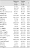
The study subjects consisted of 38 men (63.3%) and 22 women (36.7%), with a mean age of 48.4±9.6 years old. The baseline characteristics of the hypertensive patients are shown in Table 1. The clinical characteristics of the two groups were well matched except for the cfPWV, which was significantly greater in the telmisartan group than that in the valsartan group (841.2±131.0 cm/s vs. 761.1±104.4 cm/s, p<0.05).
Blood pressure control
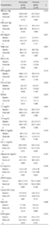
After 12 weeks of treatment, both telmisartan and valsartan significantly reduced systolic (p<0.001) and diastolic BPs (p<0.001) compared with baseline (Table 2). The mean reductions in systolic BP were 20.7±18.1 mm Hg in the telmisartan group and 22.5±17.0 mm Hg in the valsartan group. The mean reductions in diastolic BP were 16.3±13.0 mm Hg in the telmisartan group and 16.8±9.3 mm Hg in the valsartan group. No significant changes in heart rate compared with baseline were observed in either treatment.
Metabolic and inflammatory profile
A 12-week administration of telmisartan and valsartan did not result in any changes in BMI, waist circumference, HbA1c, fasting plasma glucose, fasting plasma insulin, HOMA, or adiponectin compared with baseline (Table 2). No significant changes were observed compared with baseline in the lipid profiles of the patients who had received telmisartan or valsartan, but there were trends for a reduction in total cholesterol (-4.7%, p=0.075) and triglyceride (-5.6%, p=0.058) in the telmisartan group (Table 2). Interleukin-6 was not significantly reduced in either group.
Cardiovascular indices
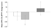
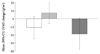
After 12-weeks of telmisartan treatment, a significant reduction in cfPWV was observed (-7.5%, p<0.05), whereas the decrease in the valsartan group was not statistically significant (Table 2). However, the results were the reverse when baseline cfPWV, which was significantly higher in the telmisartan group than in the valsartan group, was considered in the analysis. The adjusted mean {95% confidence interval (CI)} change in cfPWV from baseline was -41.8 (-87.4, 3.7) cm/s in the telmisartan group and -77.8 (-123.3, -32.3) cm/s in the valsartan group (Fig. 1). The carotid IMT and FMD did not significantly change in either group compared with baseline, although there was a trend for a reduction in the carotid IMT (-3.7%, p=0.061) in the telmisartan group (Table 2). Contrary to the slight non-significant progression (8.7%, p=0.781) of the LVMI for those patients who received valsartan, a significant regression in the LVMI (-5.9%, p<0.02) was observed in the telmisartan group at the end of the study (Table 2). But, the change in the LVMI was not significant in either group after adjusting for baseline cfPWV. The adjusted mean (95% CI) change in LVMI from baseline was -10.9 (-24.5, 2.6) g/m2 in the telmisartan group and 7.7 (-5.8, 21.3) g/m2 in the valsartan group (Fig. 2).
Differences in the changes of the study variables from baseline to the end of the study
When we compared changes between baseline and the post-treatment values in patients receiving telmisartan versus those receiving valsartan, no significant differences were observed for any of the anthropometric, hemodynamic, metabolic, or cardiovascular variables including the cfPWV (-80.5±189.2 cm/s vs. -39.1±138.6 cm/s, p=0.399), but a significant difference was observed for the LVMI. The mean changes in the LVMI were -9.6±22.7 g/m2 for the telmisartan group versus 6.4±45.3 g/m2 for the valsartan group (p<0.05). However, the mean (95% CI) differences (telmisartan group-valsartan group) in the changes of cfPWV and LVMI were 36.0 (-30.2, 102.2) cm/s (p=0.281) and -18.7 (-38.3, 1.0) g/m2 (p=0.063), respectively, both of which were not significant after adjusting for baseline cfPWV (Figs. 1 and 2). When we further adjusted BP changes and heart rate changes in addition to base-line cfPWV, the mean (95% CI) difference in the cfPWV changes, 38.7 (-30.3, 107.7) cm/s, was also not significant (p= 0.265).
Discussion
We compared the cardiovascular, metabolic, and anti-inflammatory effects of two ARB drugs, telmisartan and valsartan, in a 12-week treatment trial in patients with uncomplicated hypertension. The systolic and diastolic BPs were significantly reduced but none of the other study variables changed significantly in either group, except for cfPWV in the valsartan group. None of the changes in the measured cardio-vascular, metabolic and inflammatory parameters were significantly different between the two groups in the present study.
ARBs are preferred agents to prescribe for hypertension management in Korea16) as well as in other countries. ARBs are a valuable alternative to ACEIs in contrast to ACEIs, which many patients, particularly women, Asians, and blacks, are unable to tolerate due to adverse side-effects. ARBS are highly effective first-line agents for reducing BP as well as providing cardiovascular and renal protection, and they have the cardinal strength of improved, placebo-like tolerability and higher patient compliance rates compared with other drug classes.17)
Although ARBs share a common mechanism of action, their molecular structures and pharmacological profiles vary. However, there is a lack of large, independent, direct comparative trials for drugs within the ARB class.
Telmisartan is a compound that can simultaneously block RAS and activate PPARγ,6)7) so it has the potential to treat both the hemodynamic and biochemical features of metabolic syndrome such as hypertension, insulin resistance, and dyslipidemia. Benson et al.6) recently reported that telmisartan could function as a partial PPARγ agonist, which could influence the expression of the PPARγ target genes involved in carbohydrate and lipid metabolism, whereas telmisartan reduces glucose, insulin, and triglyceride levels in rats fed a high-fat, high-carbohydrate diet. They also reported that none of the other commercially available ARBs appeared to activate PPARγ when tested at concentrations typically achieved in the plasma with conventional oral dosing.
The ability to activate PPARγ in addition to blocking the RAS led us expect that some beneficial effects on insulin resistance, lipid profiles, adiponectin, and IL-6 would be found in the telmisartan group. But, neither telmisartan nor valsartan showed improvement in these indices. It might be because these indices are not significantly abnormal to fulfill the meta-bolic syndrome criteria at baseline in our uncomplicated hypertensive subjects and the treatment duration was too short to demonstrate significant changes. Nevertheless, trends for a reduction in total cholesterol (-4.7%, p=0.075) and triglyceride (-5.6%, p=0.058) were observed in the telmisartan group.
Telmisartan is the longest acting ARB currently available, with a mean plasma half-life of approximately 24 hours, and it has a better BP trough-to-peak ratio in the range of 0.7-1.0 in addition to having the largest distribution volume (500 L), the strongest binding affinity, and the longest duration of receptor blockade compared with other agents in this class. Telmisartan also has a rapid onset of action (maximum plasma concentrations are achieved 0.5-1.5 hours after administration).17) Compared with losartan, telmisartan shows superior efficacy during the day-time, night-time, and last 6 hours of the dosage interval.18) Moreover, using ambulatory BP monitoring, 80 mg telmisartan is superior to 160 mg valsartan in the last 6 hours of the dosing interval.19) These features of telmisartan might have caused the significant reduction in the cfPWV from baseline, which represents aortic stiffness. But the reduction was marginally insignificant after adjusting for baseline cfPWV, which was significantly higher in the telmisartan group, suggesting that the baseline level of aortic stiffness may strongly affect the changes in cfPWV caused by anti-hypertensive treatment. In contrast, the adjusted cfPWV decreased significantly from baseline in the valsartan group. Although the mean reductions in BP were not significantly different between the two groups, slightly more of a reduction (1.7 and 0.6 mm Hg, systolic and diastolic, respectively) in the valsartan group may have affected the result. Because BP is one of the strongest influencing factors of aortic stiffness,20) a reduction in cfPWV along with decreasing BP, even with a short term follow-up, was not surprising.21-23) However, the difference in the changes of the adjusted cfPWV from baseline was not significant between the two treatments (Fig. 1).
Because the aorta is the first afterload interface for the left ventricle, LVMI should be significantly affected by aortic stiffness. Therefore, we also adjusted baseline cfPWV when we compared the changes in LVMI from baseline between the two groups. The adjusted changes in LVMI were not significant in either group, and the difference in the LVMI changes between the two groups was also not significant, though it was favorable for the telmisartan group (Fig. 2). A 12-week ARB treatment may be insufficient to show a significant improvement in LVMI either by lowering the BP or by BP-independent efficacy, such as the myocardial fibrosis regression demonstrated in experimental rat models24)25) and patients with hypertension.26)
Although changes in the other vascular indices, such as the carotid IMT and FMD, were not significant in either treatment, a trend for a reduction in the carotid IMT (-3.7%, p=0.061) was observed in the telmisartan group.
This study had several potential limitations. First, the number of participating subjects was small, and the duration of treatment may have been insufficient to demonstrate overt differences between the baseline and the end of the study and between the two groups. Because this study was designed as an open-labeled pilot study to prepare for a larger and longer prospective study, we believe the results must be interpreted with caution. Second, although we performed this study with a small number of patients, we evaluated LVMI using echocardiography. Echocardiographic quantification of ventricular mass has been perceived as less reliable because of limited echocardiography standardization and because of its reliance on left ventricular wall thickness measurements and geometric assumptions about the shape of the left ventricle.27) If we had used cardiac magnetic resonance imaging, which is regarded as the current standard of reference for an accurate and reproducible in vivo measurement of left ventricular mass, the results may have been different with this small number of patients. But, we tried to reduce potential errors with repeated LVMI measurements.
We conclude that the effects of a 12-week of treatment with two ARBs, telmisartan and valsartan, on the cardiovascular, metabolic and inflammatory parameters were not significantly different in patients with uncomplicated hypertension.
 XML Download