

 PDF
PDF ePub
ePub Citation
 Print
Print


Introduction
An atrial septal defect (ASD) is a common congenital heart defect frequently detected in adulthood. Repair by transcatheter occlusion is a safe and effective method.1) The Amplatzer septal occluder (AGA medical Corp, Golden Valley, MN, USA) has been proven to be as effective as surgical closure, with the added benefit of shorter hospital stay.2) Multiple or fenestrated ASDs that require closure are not uncommon,3) and there are reports on transcatheter closure by using multiple devices.4-6) We herein report our experience with transcatheter closure of multiple ASDs by using the Amplatzer device.
Cases
Case 1
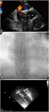
A 35 year-old woman weighing 59 kg was referred for evaluation of chest pain. The patient complained of mild exertional dyspnea. Chest radiograph revealed mild cardiomegaly. Clinically, no definite heart murmur was auscultated. Her ECG showed incomplete right bundle branch block. Two-dimensional echocardiogram showed a secundum ASD with a diameter of 17-18 mm, and a volume overloaded right ventricle. The defect was located in the anterior superior septum without a retro aortic rim. Intra-cardiac echocardiography revealed another small ASD in the posterior inferior septum, 7-8 mm in size (Fig. 1A). The distance between the two defects was 15 mm. A 24 mm and 8 mm Amplatzer septal occluder were used for transcatheter ASD closure. The two devices were subsequently implanted successfully (Fig. 1B). Cardiac CT, performed 24 hours post-intervention, showed two separate devices. Echocardiogram performed immediately after the procedure demonstrated a small leakage. Repeat echocardiogram performed six months later showed significantly reduced leakage without associated hemodynamic complications, even though the two devices were positioned on different planes (Fig. 1C). This patient was in good condition at seven-month follow up.
Case 2
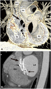
A 38 year-old male weighing 75 kg was referred for evaluation of arrhythmia. The patient was asymptomatic and physical examination revealed normal heart sounds. ECG showed benign, intermittent premature ventricular contractions. A 2D echocardiogram was performed that showed right ventricular chamber enlargement and multiple secundum ASDs, the largest one measured 12 mm, the second 8.4 mm, and the third was 5.3 mm in size. Cardiac CT showed three interatrial septal defects. The first and second ASD was partitioned by a 3.5 mm intervening septum, and the largest dimension of the first one was 13 mm without a retro aortic rim. The second was 9 mm in dimension, and the distance between the second and third ASD was 7 mm. The third ASD was 8 mm in dimension (Fig. 2A). Cardiac catheterization showed left to right shunting of 1.8, as determined by the Ficks method. There was no pulmonary hypertension. Intracardiac echocardiographic guidance was used during transcatheter closure. With some technical difficulties, a 35 mm Amplatzer cribriform occluder and a 12 mm Amplatzer septal occluder were implanted successfully. A cribriform occluder was chosen because the guide wire could not select the largest defect. Cardiac CT, performed 24 hours after implantation, showed two interleaved devices (Fig. 2B). Although a small shunt was detected on echocardiography performed immediately after the procedure, complete occlusion was demonstrated on repeat echocardiography six months later, and there were no detectible positional changes of the cribriform device.
Discussion
Transcatheter closure of ASDs has a very high success rate and very low complication rate in both children and adults.7) In Korea, Kim et al. had reported successful transcatheter closure of ASDs with the Amplatzer septal occluder (ASO), but operation was done in case of multiple ASDs.8) Multiple ASDs are detected in approximately 10% of patients with ASDs.9) Multiple defects can be closed with multiple devices with good immediate and long-term results.6) Major complications associated with ASO device include embolization and erosion. Only Awad et al.6) reported one case of embolization within 24-hours of the procedure, and one case of erosion occurred two years after the procedure. Transcatheter closure of ASDs with multiple devices has advantages over surgical closure, not only clinically but also financially in Korea. Therefore, it is a good option for the treatment of multiple ASDs.
In the first case described above, we used two ASOs. The larger defect was closed first, because the smaller defect was detected after closure of the larger defect. Most interventionists recommend smaller defects be closed first. However, Bramlet and Hoyer4) suggested that the order of closure was not crucial and regardless of the order, successful closure of all complex defects can be achieved. The second and third ASDs were easy to detect after closure of the largest defect. Many factors are considered on the order of closing multiple defects, such as the order of detection and technical difficulty.
In this case, balloon sizing was not used because of operator preference. Even though balloon sizing is a useful method to help select the appropriate device, recent studies have proposed transcatheter ASD closure without balloon sizing.10) We have performed many successful transcatheter ASD closures without balloon sizing. Awad et al.6) reported that device larger than 30-40% of the defect's 2D diameter worked well without complications. However, other clinicians have recommended balloon sizing to determine the presence of additional defects, and to formulate an appropriate closure strategy.4) In our experience, transcatheter closure without balloon sizing had shorter radiation time and lower risk of over-sizing.
In the second case, there were technical difficulties in choosing the appropriate repair order among multiple ASDs. Three ASDs were found and we tried to select the largest one for repair first. However the smallest defects were selected in all prior attempts. Therefore, the Amplatzer cribriform occluder was implanted successfully. The Amplatzer cribriform occluder is effective in covering a multiple fenestrated atrial septum. The largest cribriform device covered two of the defects successfully in this case. Another ASO was deployed for the remaining ASD. A small residual shunt was detected by color echocardiogram, 24 hours post intervention. However, 6 months later, minimal to no shunt flow was detected.
ECG gated cardiac CT is a useful method for the assessment of ASD prior to insertion of an Amplatzer device.10) To facilitate selection of device size, the defect's exact measurement is very important and therefore a good image of the defect is crucial. Cardiac CT is more useful than 2D echocardiogram in adults. In addition, cardiac CT aided visualization of the Amplatzer device at the atrial septum after implantation, especially with multiple devices. In the first case, the two devices were confirmed to be adequately separated. However, in the second case, the two devices were interconnected, as demonstrated by cardiac CT.
 XML Download