

 PDF
PDF ePub
ePub Citation
 Print
Print


Introduction
Congenital left ventricular aneurysm (CVA) is a rare clinical entity, defined as having a wide orifice connection to the ventricular cavity.1)2) The etiology is largely unknown but several theories exist, such as intrinsic abnormality during embryogenesis, infection and ischemia.3)4) The advancement of echocardiography has led to early diagnosis, including prenatal, such that several cases have been diagnosed before birth and were reported.1)3)5) We present one such case of a neonate prenatally diagnosed with a large CVA and left ventricular (LV) dysfunction, who was successfully treated with a modified Damus-Kaye-Stansel (DKS)/Dor procedure.
Case
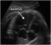
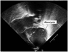
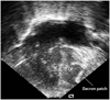
A 33-year-old woman was referred at 21 weeks' gestation with abnormal fetal echocardiographic findings, showing a large apical LV aneurysm (16.2×7.7 mm) (Fig. 1). During follow-up, LV aneurysmal dilatation had progressed (41×21 mm) with newly developed mitral regurgitation. At 33+6 weeks' gestation there was antegrade aortic flow combined with aortic regurgitation (AR). Due to progressive LV dysfunction, urgent caesarian section was performed. A female infant weighing 3.6 kg was born at 37+6 weeks' gestation. Apgar score was 2 and 6 at 1 and 5 minutes, respectively. Postnatal echocardiography showed similar findings of fetal echocardiography: a large LV apical aneurysm with maximum diameter of 41×23 mm that was larger than that of the functioning LV (Fig. 2). No visible LV forward flow was detected with moderate to severe MR. Measured LV ejection fraction (EF) was 39% and LV dimension was markedly increased (35 mm). Coronary artery origins and proximal course was normal. The aortic arch was not obstructed, but there was retrograde flow through the patent ductus arteriosus. A diagnosis was made of a large congenital LV apical aneurysm (CVA) with severe LV dysfunction (EF 39%). After considering several options, a modified DKS/Dor procedure was performed at 3 days of age. This procedure consisted of transaction of the main pulmonary artery (MPA), end-to-side anastomosis of the proximal MPA and ascending aorta, an right ventricle to pulmonary artery (RV-PA) conduit with a 6 mm Gore-tax patch graft with partial clipping, atrial septectomy and division of the patent ductus arteriosus. LV aneurysmal tissue was resected with endoventricular reductoplasty, using a Dacron patch, as it was expected to enhance ventricular geometry so that remaining LV function would be recovered. However, after cardiopulmonary bypass (CPB) weaning, intraoperative echocardiography showed a still decreased LV function and residual severe MR which was not repairable. The patient could be weaned from CPB after MV patch obliteration with high-level inotropic support in an opened sternum state. Postoperative echocardiography (Fig. 3) showed restored LV geometry with decreased LV dimension (19 mm), slightly improved LV function and good systemic and pulmonic flow from the RV. However, progressive AR with no antegrade LV flow was also detected. Finally, aortic valve obliteration with delayed sternal closure was performed on postoperative day 14. Histology on the resected LV myocardium showed myocardial hypertrophy with multiple fibrotic scars. She was discharged on postoperative day 43 with diuretics medication. During follow-up, progressive cyanosis was noted. Echocardiography and cardiac computed tomography showed RV-PA conduit narrowing with peak velocity of 4.6 m/sec. It was decided that a bidirectional cavopulmonary shunt with RV-PA conduit division would be performed at 6 months of age, which occurred without negative sequelae. The patient is currently 21 months old and in good general health.
Discussion
CVA is a rare cardiac abnormality. It has a wide communication to the LV which differs from congenital diverticulum of the heart, which is characterized by a finger-like appendix emerging from the wall of the LV.2)6) Usually, both are found in the apex or paravalvular area, and thus correction remains sometimes challenging. Early accurate diagnosis is important to predict the prognosis and manage the problem. The etiology and natural history are not clearly understood, though several factors were considered as possible causes, including infection, ischemic insult or developmental or genetic abnormality, based on the reports of cases which was diagnosed during prenatal period.7)8)
The prognosis is variable, depending on the size, timing of diagnosis, growth and signs of cardiac failure. Depending on the amount of myocardial fibers involved, aneurysm shows almost normal contractility and can be dyskinetic or akinetic.2) Previously reported, large aneurysm were often associated with poor prognosis.9-11)
In this case, she was diagnosed early with a large CVA, in the prenatal period at 21 weeks of gestation. Serial fetal and postnatal echocardiography showed progressive aneurysmal enlargement and ventricular dysfunction.
A treatment strategy for CVA has not been clearly established due to the small number of published cases to go on. Therefore, treatment should be individually tailored depending on clinical severity, which can vary from asymptomatic to arrhythmias, severe fetal hydrops, pericardial effusion, heart failure, peripheral embolism, and fetal demise.2)8)11)13-17) There appears to be a consensus for surgical intervention in severely symptomatic patients1)2) and many authors also agree even in asymptomatic patients to prevent serious complications.
Several surgical options were reported, such as aneurysmectomy alone or combined with mitral valve repair and pericardial or Dacron patch closure of LV connection site.8)16)
It was reported that partial left ventriculectomy in neonates with dilated cardiomyopathy resulted in successful LV remodeling and recovered LV function.14)
In the present case, she was severely symptomatic with too large a CVA to permit proper LV function. Because the LV did not contribute to systemic perfusion, we diagnosed it as functional hypoplastic left heart syndrome and decided to perform uni-ventricular palliation using a modified DKS procedure in addition to combined Dor procedure. Through this we expected improved LV geometry, which would contribute to recovery of LV function. However, intra-operative evaluation showed that residual mitral valve regurgitation was too severe to be repaired, which was an aforementioned combined morphologic abnormality.10) Therefore, we had no choice but to perform left sided valve obliteration.
The Norwood/Batista operation was previously used on an infant with dilated cardiomyopathy,18) however, to our knowledge, it had not been applied in an infant with CVA and severe LV dysfunction.
In summary, CVA is a rare entity with variable outcomes. Early diagnosis during the prenatal period with large sized, progressive CVA was an indicator of poor prognosis. An individualized therapeutic approach is recommended, depending on the clinical presentation. In severe cases with LV dysfunction, such as the case of the presented patient, surgical intervention is recommended. A modified DKS/Dor procedure can be considered as a possible palliation with a good result.
 XML Download