Abstract
Aortic rupture has a high mortality rate and can be considered a medical emergency. The standard treatment for aortic rupture is surgical repair. An aortic stent graft for a ruptured descending aorta is considered an effective alternative treatment. However, an aortic stent graft is difficult when the aortic aneurysm is in the aortic arch due to supra-aortic vessels. We report on a patient with a ruptured aortic arch aneurysm treated with a hybrid procedure, which involved a carotid to carotid bypass operation and an aortic stent graft. A 71-year-old male patient visited our cardiovascular center suffering from hemoptysis. The chest CT and aortography showed a 9 cm sized aortic arch aneurysm 0.5 cm distal to the left subclavian artery and a hemothorax in the left lung. The patient refused to undergo a full open operation. We performed a carotid to carotid bypass in advance, and two pieces of aortic stent grafts were placed across the left carotid artery and left subclavian artery. The follow up CT showed the aortic stent grafts, no endoleaks and no thrombus in the aortic arch aneurysm. The patient was discharged from the hospital without complication.
The rupture of a thoracic aortic aneurysm can be a life-threatening condition. The risk of rupture is highly associated with the aneurysm size. The estimated 5 year risk of rupture of an aneurysm with a diameter between 4 and 5.9 cm is 16%, but it rises to 31% for aneurysms greater than 6 cm.1) Surgical repair has been considered the preferred method of treatment for many years, but the postoperative mortality and morbidity rates still remain high.2) Surgical repair is also technically difficult because a surgical approach to the descending aorta via the left thorax and lung is often complicated. Endovascular aortic stent graft placement has recently been introduced for repairing descending thoracic aneurysms and is widely regarded as an emerging alternative option to surgery in patients with serious coexisting illnesses.
The endovascular technique is minimally invasive and is associated with less mortality and morbidity.3) The endovascular deployment of stent grafts for treating descending thoracic aortic aneurysms has been shown to be clinically feasible. Previous studies have shown that the descending thoracic aorta represented an easy area for deployment,4) whereas this procedure for an aortic arch aneurysm is more difficult because of the curvature of the aortic arch and the presence of the supra-aortic vessels.5) The technical improvement with more flexible stent grafts and better revascularization procedures has recently allowed deployment of stent grafts in the aortic arch in most cases.6) Here, we report on a patient with a ruptured aortic arch aneurysm where the patient was successfully treated by an aortic stent graft with no complications.
A 71-year-old male patient with diabetes mellitus and hypertension visited the emergency room of our hospital because of hemoptysis. He was diagnosed with a 7 cm sized aortic arch aneurysm 2 years earlier. He was recommended to undergo an operation for the aneurysm, but refused to do so. He also did not take his medicine and was without a follow up examination over that period.
At the time of admission, his blood pressure was 150/100 mmHg, pulse rate was 86 beats/min, respiratory rate was 20 breaths/min and body temperature was 36.5℃. The patient was alert, but looked quite ill in appearance. His heart sounds were regular without murmurs, and the lung sounds were decreased at the left lung field. All other aspects of the physical examination were normal.
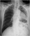
The initial laboratory findings showed that his hemoglobin level was 8.1 g/dL, white blood cell count was 9.7×10³/µL and platelet count was 170×103/µL. Additional laboratory studies were normal. An electrocardiogram showed normal sinus rhythm without specific abnormal findings and a chest X-ray showed a mass-like lesion in the left hilar zone (Fig. 1). The chest CT showed a 9 cm sized saccular aneurysm 0.5 cm distal to the left subclavian artery with thrombus in the aortic arch, which was oozing to the anterior wall owing to rupture of the aneurysm, and there was a hemothorax in the left lung (Fig. 2). Labetalol was infused intravenously for controlling blood pressure.
We recommended an operation for treating the ruptured aortic arch aneurysm, but the patient and his family refused an operation because of the risk of surgery. Therefore, we decided to perform a hybrid procedure that consisted of a carotid to carotid bypass operation and the placement of stent grafts in the aortic arch aneurysm to save the left carotid artery. We planned to sacrifice the left subclavian artery intentionally and later performed a second stage left carotid to left subclavian artery bypass operation if the patient displayed subclavian steal syndrome, severe dizziness and/or left arm pain. Before endovascular stent graft placement, we checked the whole aortogram and excluded the presence of stenosis that would interfere with collateralization in the carotid or vertebral arteries. We referred him to a thoracic surgery to perform the carotid to carotid bypass operation connecting from the right carotid artery to the left carotid artery in advance for revascularization of the post-stent grafting. Insertion of the aortic stent grafts was performed following the successful by-pass operation. The aortography showed the presence of a huge aortic arch aneurysm 0.2 cm distal to the left subclavian artery (Fig. 3A). We deployed two pieces of aortic stent grafts (40 mm×10 cm, 36 mm×10 cm, S&G biotech, Korea) across the left carotid artery and left subclavian artery through the left femoral artery (Fig. 3B, C, and D). The second aortic stent graft (36 mm×10 cm, S&G biotech) was deployed in the first aortic stent graft with a 7 cm overlapped segment to protect against disconnection of the aortic stent grafts. The post-procedure aortography showed no endoleak or other complications.
Directly after the procedure, the patient was transferred to the intensive care unit, where he complained of aggravated dyspnea and massive hemoptysis. Chest X-ray showed total haziness in the left lung field and a chest CT showed successful stenting in the aortic arch aneurysm and total ateletasis of the left lung due to external compression of the left main bronchus by hematoma (Fig. 4). Therapeutic bronchoscopy was performed to remove a large amount of blood clots in the left bronchus. The left atelectasis improved on the chest X-ray taken 5 days after a bronchoscopy procedure (Fig. 5A). Follow up CT angiography of the aorta after 7 days showed successful stenting, no endoleak and no thrombus in the aortic arch (Fig. 5B and C).
The patient was discharged from our hospital without any further adverse clinical events. No cardiovascular events were observed during 1 year of follow-up.
The incidence rate of thoracic aortic aneurysms is 10.4 per 100,000 person per year, and has recently been reported to be increasing.1) Some of the causes of thoracic aortic aneurysms include cystic medial necrosis, atherosclerosis, Marfan syndrome and a bicuspid aortic valve. Most thoracic aortic aneurysms are asymptomatic. However, compression of the surrounding tissue by aneurysm may cause hoarseness, dyspnea, chest pain and/or dysphagia.7) Rupture of a thoracic aortic aneurysm is the most important complication because it is a life-threatening condition. The risk of rupture is highly associated with the size of the aneurysm. The 5 year risk of rupture is 0% for aneurysms less than 4 cm in diameter, 16% for aneurysms that are 4 to 5.9 cm, 31% for aneurysms 6 cm or larger and the cumulative risk is 20% after 5 years.1) Rupture of a thoracic aortic aneurysm represents 32-47% of the causes of death in patients with thoracic aortic aneurysms.8)
Operative repairs with placement of a prosthetic graft are regarded as the standard treatment for a ruptured thoracic aortic aneurysm. However, ruptured thoracic aneurysms complicated by perforation have a surgical mortality rate that exceeds 50% despite advances in operative procedures and postoperative care.9) Surgery related deaths are caused by myocardial infarction, heart failure, cerebral infarction, renal failure, bleeding and sepsis. These deaths increase in patients who have underlying coronary heart disease and heart failure, or are old in age.8) Open surgical repair is technically difficult because the surgical approach to the descending aorta via the left thorax and lung is complicated. Endovascular therapy by deployment of stent grafts has recently been shown to be a feasible technique for the treatment of ruptured thoracic aneurysms. Considering the short time of the procedure and minimally invasive approach of the endovascular technique, it has been suggested as an elective or emergency procedure for a wide spectrum of iatrogenic conditions.10)
It is often necessary to use the segment between the left common carotid artery and the left subclavian artery as the proximal landing zone with subsequent coverage of the left subclavian artery for the safe fixation of stent grafts in the distal aortic arch. Intentional coverage of the left subclavian artery carries a low risk according to clinical experiences, if stenoses and abnormalities of the supra-aortic and intracranial arteries supplying the brain are ruled out11) and prophylactic subclavian-carotid transposition or extrathoracic bypass grafting has reduced these risks.12) Accurate placement in the arch becomes more challenging by the high blood flow and substantial movement of the arch with each heartbeat. Intravenous nitroprusside, nicardipine and nitroglycerine are used to reduce blood pressure during the deployment of a stent graft. New materials with better flexibility, precise delivery, reliable fixation and increased durability have recently been introduced and this allows deploying in the arch in most instances.
In the present case, a huge aortic arch aneurysm originated just beyond the left subclavian artery, and coverage of both the left subclavian artery and the left common carotid artery orifices was inevitable and placement of long stent grafts was needed for their safe fixation. Before endovascular stent graft placement, we checked the whole aortogram and excluded the presence of stenosis that would interfere with collateralization in the carotid or vertebral arteries. Since we accepted the results of previous studies that the possible arm ischemia was mostly mild and could be managed secondarily, we planned intentional coverage of the left subclavian ostium. Next, we proceeded with the operation, connecting from the right carotid artery to the left carotid artery for revascularization of the post-stent grafting because vertebral territory stroke after left subclavian artery coverage is indeed a documented risk when the left common carotid artery orifice is intentionally covered. Although there was a high risk of mortality due to the patient having a poor general medical condition and unexpected emergency situation, the results of endovascular repair were satisfactory.
The massive hemoptysis of the patient was thought to be caused by an aortopulmonary or aortobronchial fistula. Both are unusual complications associated with aortic aneurysm and usually occur after erosion or rupture of a degenerative or false aneurysm of the distal aortic arch.13)14) Since the patient had no pre-existing pulmonary disease, surgical history and hemoptysis, an aortopulmonary fistula might have formed due to erosion from the continuous pulsatile friction between the pulmonary artery and the aortic aneurysm wall, or an aortobronchial fistula between the aortic and bronchial lumina was created because of compression of the tracheobronchial tree by the enlargement of the thoracic aneurysm. This may have induced pressure necrosis, which can lead to erosion of both the aortic and bronchial walls.15)16) Endovascular stent graft repair of an aortopulmonary fistula appears to be safe and well tolerated.17)
In conclusion, a hybrid procedure with a right to left common carotid artery bypass operation and an aortic stent graft for the treatment of aortic arch aneurysm can be a beneficial alternative to surgical treatment, especially for patients with co-existing illnesses.
Figures and Tables
 | Fig. 2The chest CT scan shows a saccular aneurysm with intramural thrombus in the aortic arch (A). The aortic aneurysm is suspected to be leaking to the anterior chest wall (white arrow) (B) and the left main bronchus is compressed by the huge aortic aneurysm (black arrow) (C). |
 | Fig. 3Aortography shows a huge aortic arch aneurysm 0.2 cm distal to the left subclavian artery (A) and the process of deploying 2 pieces of aortic stent grafts (40 mm×10 cm, 36 mm×10 cm, S&G biotech, Korea) across the left carotid artery and left subclavian artery (B, C and D). |
 | Fig. 4The chest X-ray shows total atelectasis of the left lung (A). The left main bronchus is compressed due to the posteriorly displaced left pulmonary artery, resulting in left lung total atelectasis on the chest CT scan (black arrow) (B) and there is no endoleak or occlusion of the neck vessels (C). |
References
1. Clouse WD, Hallett JW Jr, Schaff HV, Gayari MM, Ilstrup DM, Melton LJ 3rd. Improved prognosis of thoracic aortic aneurysm: a population-based study. JAMA. 1998. 280:1926–1929.
2. Kouchoukos NT, Dougenis D. Surgery of the thoracic aorta. N Engl J Med. 1997. 336:1876–1888.
3. Dake MD. Endovascular stent-graft management of thoracic aortic diseases. Eur J Radiol. 2001. 39:42–49.
4. Dake MD, Miller DC, Semba CP, Mitchell RS, Walker PJ, Liddell RP. Transluminal placement of endovascular stent-grafts for the treatment of descending thoracic aortic aneurysms. N Engl J Med. 1994. 331:1729–1734.
5. Criado FJ, Barnatan MF, Rizk Y, Clark NS, Wang CF. Technical st-rategies to expand stent-graft applicability in the aortic arch and pro-ximal descending thoracic aorta. J Endovasc Ther. 2002. 9:Suppl 2. II32–II38.
6. Czerny M, Fleck T, Zimpfer D, et al. Combined repair of an aortic arch aneurysm by sequential transposition of the supra-aortic branches and endovascular stent-graft placement. J Thorac Cardiovasc Surg. 2003. 126:916–918.
7. Halperin JL, Olin JW. Fuster V, editor. Disease of the aorta. Hurst's The Heart. 2004. 11th ed. New York: McGraw-Hil;2304.
8. Cohn PF, Braunwald E. Braunwald E, editor. Disease of the aorta. Heart Disease. 1997. 5th ed. Philadelphia: Saunders;1550.
9. Johansson G, Markstrom U, Swedenborg J. Ruptured thoracic aortic aneurysms: a study of incidence and mortality rates. J Vasc Surg. 1995. 21:985–988.
10. Sunder-Plassmann L, Scharrer-Pamler R, Liewald F, Kapfer X, Gorich J, Orend KH. Endovascular exclusion of thoracic arotic aneurysm: mid-term results of elective treatment and in contained rupture. J Card Surg. 2003. 18:367–374.
11. Hausegger KA, Oberwaldner P, Tiesenhausen K, et al. Intentional left subclavian artery occlusion by thoracic aortic stent-grafts without surgical transposition. J Endovasc Ther. 2001. 8:472–476.
12. Zipfel B, Buz S, Hammerschmidt R, Hetzer R. Occlusion of the left subclavian artery with stent grafts is safer with protective reconstruction. Ann Thorac Surg. 2009. 88:498–505.
13. Killen DA, Muehlebach GF, Wathanacharoen S. Aortopulmonary fistula. South Med J. 2000. 93:195–198.
14. Favre J, Gournier J, Adham M, Rosset E, Barral X. Aortobronchial fistula: report of three cases and review of the literature. Surgery. 1994. 115:264–270.
15. Kim SE, Kim HJ, Lee SH, et al. A case of aortopulmonary fistula caused by a huge thoracic aortic aneurysm. Korean Circ J. 2009. 39:209–212.
16. Nishizawa J, Matsumoto M, Sugita T, et al. Surgical treatment of five patients with aortobronchial fistula in the aortic arch. Ann Thorac Surg. 2004. 77:1821–1823.
17. Kochi K, Okada K, Watari M, Orihashi K, Sueda T. Hybrid endovascu-lar stent grafting for aortic arch aneurysm with aortopulmonary fistula. J Thorac Cardiovasc Surg. 2002. 123:363–364.


 PDF
PDF ePub
ePub Citation
 Print
Print


 XML Download