Abstract
Background and Objectives
Reports on the incidence of intracardiac thrombi (ICT) have increased over the last few decades, but ICT are still relatively rare among children. Left ventricular systolic dysfunction and dilatation may contribute to the formation of ICT, especially when a hypercoagulable state exists. The aim of this study was to describe the incidence of ICT in children suffering from cardiac failure with left ventricular dysfunction and to identify risk factors on admission for developing ICT.
Subjects and Methods
We conducted a retrospective chart review of children up to 18 years of age admitted to the Pediatric Intensive Care Unit due to cardiac failure with left ventricular dysfunction between January 1, 2003 and December 31, 2008.
Results
Twenty-one patients were admitted with clinical signs of cardiac failure and echocardiographic findings compatible with dilated cardiomyopathy or acute myocarditis. Dilated cardiomyopathy was diagnosed in 11 patients (52%). Adenoviruses and enteroviruses were suspected to be the cause of acute myocarditis in 5 cases. The personal or family history of hypercoagulable states were obtained from 19 out of 21 patients (90%). Among patients with a hypercoagulable state, 3 out of 7 developed ICT compared with none out of 12 among patients without hypercoagulability (p=0.043). Two of these 3 patients experienced an embolic event.
Intracardiac thrombi (ICT) are relatively rare among children. Reports on the incidence of ICT have increased in the last few decades, especially when a hypercoagulable state exists.1-5) Left ventricular systolic dysfunction and dilatation may contribute to the formation of ICT by altering the hemostatic state with concomitant blood stasis within a dilated left ventricle. Furthermore, heart failure is associated with abnormalities of endothelial function.6-8) In patients with congenital heart diseases, causes of ICT may differ, depending on the chamber of the heart involved.
Thrombi of the right side are usually associated with the presence of central venous catheters, endocarditis and polycythemia, 9)10) while dilated cardiomyopathy, atrial fibrillation, endocarditis, prosthetic heart valves, intracardiac tumors and rheumatic mitral stenosis are the most important predisposing factors for cardiac emboli sources when the left chamber is involved. A significant complication of ICT is the development of cerebral thrombosis.11) Recently, reports of ICT as an important cause of mortality in post-Fontan procedure patients are increasing.12)
We hereby present our experience in pediatric patients who presented with clinical cardiac failure due to acute myocarditis or dilated cardiomyopathy and developed secondary complications of the left ventricular thrombi.
This study was approved by the Institutional Review Board of our Medical Center. We retrospectively reviewed the medical records of all patients up to 18 years of age admitted to the seven-bed pediatric intensive care unit (PICU) of a general hospital from January 1, 2003 to December 31, 2008 due to acute myocarditis or dilated cardiomyopathy accompanied by a low systolic ejection fraction of below 40%. Patients were identified using the hospital computerized database. Those in a state of pre-shock or shock due to non cardiogenic causes were excluded.
All pediatric patients with clinical signs of left heart failure, an enlarged heart shadow on chest radiographs, and left ventricular dysfunction with an ejection fraction below 40% on two-dimensional echocardiography as measured by Simpson's method13) were included. The selection of a 40% ejection fraction was based on the American Society of Echocardiography guidelines that define an abnormal ejection fraction as <55%, with the cutoffs for moderately abnormal and severely abnormal at 44% and 30%, respectively.14) The diagnosis of dilated cardiomyopathy was based on definitions and classifications of cardiomyopathies by the American Heart Association in 2006, ventricular chamber enlargement and systolic dysfunction with normal left ventricle wall thickness, as determined by two-dimensional echocardiography.15) Grading of mitral regurgitation was estimated by color flow Doppler echocardiography. Acute myocarditis was diagnosed on the basis of clinical and laboratory results. Polymerase chain reaction (PCR) of nasopharyngeal and rectal swabs was performed in patients with a history of acute viral disease that preceded cardiac failure such as upper respiratory tract infection, diarrhea and fever.
Thrombophilia workup including prothrombin time, activated partial thromboplastin, protein C, protein S, antithrombin III, activated protein C resistance, thrombophilic mutations of factor V Leiden and thermolabile methylenetetrahydrofolate reductase (MTHFR C677T), prothrombin G20210A, antiphospholipid antibodies and plasma level of homocysteine was performed in the 3 patients that developed ICT.
Data was extracted on patient demographics, mode of presentation, clinical features, investigation on admission, inpatient management and follow up. Patients with ICT were characterized and compared to the rest of the group. The incidence of ICT in patients with and without hypercoagulability was compared by the Fisher Exact test.
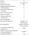
Twenty-one patients were admitted to the PICU due to cardiac failure with acute myocarditis or dilated cardiomyopathy. All patients had clinical signs of cardiac failure on admission. A chest X-ray revealed a cardiac shadow enlargement in all the participants and signs of lung congestion in 17 patients (81%). The characteristics of the patients are summarized in Table 1. Anamnesis details of a personal or family history predisposing the patient to hypercoagulability were obtained in 19 patients (90%). All patients underwent echocardiography within 24 hours of admission, which revealed heart enlargement, varying degrees of mitral regurgiation and a decreased ejection fraction. Echocardiography was repeated at intervals of 48 to 96 hours. Dilated cardiomyopathy was diagnosed in 11 (52%) patients. Adenoviruses and enteroviruses were suspected to be the cause of acute myocarditis in 5 cases.
A family history of thrombophilia was found in 5 patients (deep vein thrombosis, stroke, recurrent abortions), and a personal high risk for a hypercoagulable state was found in 2 patients (signet cell carcinoma, long flight). Within 24 hours of admission, low-molecular-weight heparin (LMWH) was administered in 14 patients (66%). All patients with a personal or familial high risk of thrombophilia were treated with LMWH on admission.
According to our policy, every child admitted with moderate to severe left ventricle dysfunction was treated with LMWH. Some of the patients were not treated with LMWH due to lack of awareness of the care team.
ICT were observed in 3 (14%) patients, all of whom had sinus rhythm. Among the patients with a hypercoagulable state, 3 out of 7 developed ICT compared with none out of 12 among patients without hypercoagulability (p=0.043). The characteristics of the patients with ICT are summarized in Table 2.
Patient No. 1 (Table 2) had a family history of stroke and was treated with LMWH within 24 hours of admission as a prophylactic dose, but developed ICT 48 hours later. Work up for thrombophilia revealed the MTHFR C677T mutation. Patient No. 2 (Table 2) was also treated with LMWH within 24 hours of admission as a prophylactic dose, due to a hypercoagulable state (signet cell carcinoma), but developed ICT on day 5 after admission during a chemotherapy course of 5-fluorouracil (5-FU). When the treatment with 5-FU was stopped, rapid cardiac improvement occurred. Work up for thrombophilia revealed heterozygosity for the MTHFR C677T mutation. Patient No. 3 (Table 2) had a known dilated cardiomyopathy that had been controlled by conservative medication for a period of 9 months. One week prior to admission, she stopped taking her medication and flew from Peru to Israel. This patient already had ICT on admission that probably developed after a long-haul flight from Peru to Israel.24) Thrombophilia work up revealed normal results. Two of these patients developed embolic events, 1 of them involving the brain. The mean ejection fraction was 18.6% in patients with ICT compared with 18% in patients without ICT
Five patients required mechanical ventilation on admission, and 3 of them deceased (signet cell carcinoma, mitochondrial disease, and very long chain acyl-CoA dehydrogenase deficiency). All 21 patients were followed up for a median period of 14 months (range of 29 months) and in 3 of these patients normal heart function was restored with suspected acute myocarditis.
Of the 5 patients with a positive familial history of hypercoagulability, 1 suffered from suspected acute myocarditis due to enterovirus, 1 suffered from anomalous origin of coronary artery (AOCA) and the other 3 had idiopathic dilated cardiomyopathy. Four of them were treated with LMWH on admission (AOCA excluded). Heart function was completely restored in the patient with acute myocarditis, improved significantly in the patient with AOCA and remained poor in the rest.
In a cohort of 21 pediatric patients admitted for acute myocarditis or dilated cardiomyopathy with a significant decrease in ejection fraction, 3 (14%) suffered from ICT. A similar incidence (14%) was reported by Günthard et al.16) when describing 130 pediatric patients with dilated cardiomyopathy. In this series, patients with ICT had a seriously impaired fractional shortening (10±3%) as compared to those without thromboembolism (17±6%). Contrary to these findings, we did not find significant differences in ejection fraction between patients who did or did not develop ICT.
A personal or family predisposition to hypercoagulability was identified in 36.8% of the participants and in all patients with ICT. This finding may help to identify patients that are likely to develop ICT. The association between ICT and a hypercoagulability state is in agreement with previous studies on pediatric populations that focused on hypercoagulability states as a predisposing factor for the formation of ICT.5)6)17)18) A retrospective study was conducted that analyzed the features and risk factors of childhood thrombotic events in 59 patients with cardiac defects. Cardiomyopathy was found to be one of the risk factors for developing ICT, and 23 of the 59 described patients had at least one thrombophilic mutation.19)
In the current study, Patient No. 1 (Table 2) had a family history of stroke and was treated with LMWH as a prophylactic dose, but developed ICT. Work up for thrombophilia revealed the MTHFR C677T mutation. Patient No. 2 (Table 2) was also treated with LMWH, due to a hypercoagulable state (signet cell carcinoma), but developed ICT during a chemotherapy course of 5-FU. When the treatment with 5-FU was stopped, rapid cardiac improvement occurred. Work up for thrombophilia revealed heterozygosity for the MTHFR C677T mutation.
In the literature, 5-FU therapy in combination with other chemotherapy drugs was found to be a possible cause of angina, dysrhythmias and dilated cardiomyopathy. In some cases, the pathological findings were compatible with acute myocarditis or toxic myopathy.20-23) Cancer is one of the most important acquired risk factors for the development of thromboembolism. Tumors can express procoagulant proteins and may induce the production of inflammatory cytokines that indirectly contribute to the development of hypercoagulability.24)
Patient No. 3 (Table 2) had a known dilated cardiomyopathy that had been controlled by conservative medications for a period of 9 months. One week prior to admission, the patient stopped taking her medication and flew from Peru to Israel. A long-haul flight is a risk factor that may activate the coagulation system.25)
Results of several adult studies regarding the degree of mitral regurgitation and ICT formation suggest that severe mitral regurgitation in patients with ventricular dysfunction prevents left ventricular thrombus formation by decreasing stasis and sluggish blood flow.26) In the current study, all the patients had mitral regurgitation, but none of the 3 patients that developed ICT had severe mitral regurgitation.
It is recommended to treat adult patients with dilated cardiomyopathy who are considered to be at high risk for thromboembolism with prophylactic anticoagulants.27) In the pediatric population, there is no apparent evidence to support prophylactic anticoagulant treatment of patients with dilated cardiomyopathy. However, there is a recommendation for the routine use of anticoagulants in children with poor ventricular function within the context of dilated cardiomyopathy.16)
Unfractionated heparin or LMWH is usually the recommended anti-thrombotic therapy, unless there is a major vessel occlusion causing critical compromise of organs or limbs.28) In the current study, ICT developed despite prophylactic administration of LMWH on admission and regardless of the degree of the ejection fraction, which was similar in both groups. This emphasizes the necessity for repeated echocardiographs even when LMWH is administered, especially when a personal or familial hypercoagulable state coexists. It is unclear whether or not more aggressive anti-coagulation treatment could have prevented the thromboembolic events.
This study is limited by its retrospective nature, the small group of patients and the fact that thrombophilia work up was not performed in all participants. Furthermore, no consistent policy to prevent the formation of ICT was used in all participants and history details regarding a personal or familial history of hypercoagulability were missing in 2 patients. With the knowledge of these limitations, unequivocal conclusions cannot be reached, but we suggest taking note of specific details about personal and familial hypercoagulable states. Patients with a higher risk for developing ICT must be followed carefully when presenting with left ventricle dysfunction even when prophylactic anti-coagulation treatment has been administered. We also suggest considering thrombophilia work-up in patients with dilated cardiomyopathy and left ventricle dysfunction. Further studies are needed to determine the optimal LMWH dosage in similar cases.
Figures and Tables
References
1. Atalay S, Akar N, Tutar HE, Yilmaz E. Factor V 1691 G-A mutation in children with intracardiac thrombosis: a prospective study. Acta Paediatr. 2002. 91:168–171.
2. Atalay S, Imamoglu A, Ikizler C, Uluoglu O, Ocal B. Mitral valve and left ventricular thrombi in an infant with acquired protein C deficiency. Angiology. 1995. 46:87–90.
3. Favara BE, Franciosi RA, Butterfield LJ. Disseminated intravascular and cardiac thrombosis of the neonate. Am J Dis Child. 1974. 127:197–204.
4. Gurgey A, Ozyurek E, Gümrük F, et al. Thrombosis in children with cardiac pathology: frequency of factor V Leiden and prothrombin G20210A mutations. Pediatr Cardiol. 2003. 24:244–248.
5. Ozkutlu S, Ozbarlas N, Ozme S, Saraclar M, Gögüs S, Demircin M. Intracardiac thrombosis diagnosed by echocardiography in childhood: predisposing and etiological factors. Int J Cardiol. 1993. 40:251–256.
6. Lip GY, Gibbs CR. Does heart failure confer a hypercoagulable state? Virchow's triade revisited. J Am Coll Cardiol. 1999. 33:1424–1426.
7. Meltzer RS, Visser CA, Fuster V. Intracardiac thrombi and systemic embolization. Ann Intern Med. 1986. 104:689–698.
8. Treasure CB, Vita JA, Cox DA, et al. Endothelium-dependent dilation of coronary microvasculature is impaired in dilated cardiomyopathy. Circulation. 1990. 81:772–779.
9. Kádár K, Hartyánszky I, Király L, Bendig L. Right heart thrombus in infants and children. Pediatr Cardiol. 1991. 12:24–27.
10. Ross P Jr, Ehrenkranz R, Kleinman CS, Seashore JH. Thrombus associated with central venous catheters in infants and children. J Pediatr Surg. 1989. 24:253–256.
11. Tegeler CH, Downes TR. Thrombosis and the heart. Semin Neurol. 1991. 11:339–352.
12. Khairy P, Fernandes SM, Mayer JE Jr, et al. Long-term survival, modes of death, and predictors of mortality in patients with Fontan surgery. Circulation. 2008. 117:85–92.
13. Schiller NB, Shah PM, Crawford M, et al. American Society of Echocardiography Committee on Standards. Subcommittee on Quantitation of Two-Dimensional Echocardiograms. Recommendations for quantitation of the left ventricle by two-dimensional echocardiography. J Am Soc Echocardiogr. 1989. 2:358–367.
14. Lang RM, Bierig M, Devereux RB, et al. Recommendations for chamber quantification: a report from the American Society of Echocardiography's Guidelines and Standards Committee and the Chamber Quantification Writing Group, developed in conjunction with the European Association of Echocardiography, a branch of the European Society of Cardiology. J Am Soc Echocardiogr. 2005. 18:1440–1463.
15. Maron BJ, Towbin JA, Thiene G, et al. Contemporary definitions and classification of the cardiomyopathies: an American Heart Association Scientific Statement from the Council on Clinical Cardiology, Heart Failure and Transplantation Committee; Quality of Care and Outcomes Research and Functional Genomics and Translational Biology Interdisciplinary Working Groups; and Council on Epidemiology and Prevention. Circulation. 2006. 113:1807–1816.
16. Günthard J, Stocker F, Bolz D, et al. Dilated cardiomyopathy and thrombo-embolism. Eur J Pediatr. 1997. 156:3–6.
17. Kohlhase B, Vielhaber H, Kehl HG, Kececioglu D, Koch HG, Nowak-Göttl U. Thromboembolism and resistance of activated protein C in children with underlying cardiac disease. J Pediatr. 1996. 129:677–679.
18. Suskan E, Kemahli S, Atalay S, Ertogan F, Karademir S. Intracardiac thrombosis associated with acquired protein C deficiency. Eur J Pediatr. 1994. 153:862–863.
19. Alioglu B, Avci Z, Tokel K, Atac FB, Ozbek N. Thrombosis in children with cardiac pathology: analysis of acquired and inherited risk factors. Blood Coagul Fibrinolysis. 2008. 19:294–304.
20. Hochster H, Wasserheit C, Speyer J. Cardiotoxicity and cardioprotection during chemotherapy. Curr Opin Oncol. 1995. 7:304–309.
21. Martin M, Diaz-Rubio E, Furió V, Blázquez J, Almenarez J, Farina J. Lethal cardiac toxicity after cisplatin and 5-fluorouracil chemotherapy. Report of a case with necropsy study. Am J Clin Oncol. 1989. 12:229–234.
22. Misset B, Escudier B, Leclercq B, Rivara D, Rougier P, Nitenberg G. Acute myocardiotoxicity during 5-fluorouracil therapy. Intensive Care Med. 1990. 16:210–211.
23. Sasson Z, Morgan CD, Wang B, Thomas G, MacKenzie B, Platts ME. 5-Fluorouracil related toxic myocarditis: case reports and pathological confirmation. Can J Cardiol. 1994. 10:861–864.
24. Blom JW, Doggen CJ, Osanto S, Rosendaal FR. Malignancies, prothrombotic mutations, and the risk of venous thrombosis. JAMA. 2005. 293:715–722.
25. Schreijer AJ, Cannegieter SC, Meijers JC, Middeldorp S, Büller HR, Rosendaal FR. Activation of coagulation system during air travel: a crossover study. Lancet. 2006. 367:832–838.
26. Blondheim DS, Jacobs LE, Kotler MN, Costacurta GA, Parry WR. Dilated cardiomyopathy with mitral regurgitation: decreased survival despite a low frequency of left ventricular thrombus. Am Heart J. 1991. 122:763–771.
27. Ripley TL, Nutescu E. Anticoagulation in patients with heart failure and normal sinus rhythm. Am J Health Syst Pharm. 2009. 66:134–141.
28. Monagle P, Chan A, Chalmers E, et al. Antithrombotic therapy in neonates and children: American College of Chest Physicians Evidence-Based Clinical Practice Guidelines (8th edition). Chest. 2008. 133:6 Suppl. 887S–968S.


 PDF
PDF ePub
ePub Citation
 Print
Print


 XML Download