

 PDF
PDF ePub
ePub Citation
 Print
Print


Introduction
Vasospasm of the left main coronary artery (LMCA) is a very rare, sometimes life-threatening cardiovascular event,1)2) which may be caused by spontaneous simultaneous coronary spasm (as with variant angina in an uncommon case) or caused by an iatrogenic (catheter- or guidewire-induced) component. Although catheter-induced spasm of the LMCA has been documented,3) routine diagnostic catheterization rarely causes LMCA spasm.4) We report a case of spontaneous spasm of the LMCA detected by 64-slice multi-detector computed tomographic coronary angiography (MDCT-CA).
Case
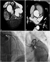
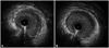
A 61-year-old male was sent to undergo coronary angiography for suspected angina from significant ostial stenosis of the LMCA found on MDCT (Fig. 1A and B). He had suffered from frequent morning chest tightness that occurred at rest during the past 2 months. He had no risk factors for coronary artery disease and did not smoke or drink alcohol. Routine laboratory findings, including a thyroid function test, were within normal limits. Significant narrowing of the LMCA compatible with the MDCT finding was detected on coronary angiography (Fig. 1C). We conducted an intravascular ultrasound (IVUS) to confirm the coronary angiographic findings after intracoronary administration of 200 µg nitroglycerine. IVUS results showed negative remodeling of the LMCA without significant atherosclerotic change (Fig. 2A). After additional administration of 500 µg nitroglycerine, we again performed coronary angiography (Fig. 1D) and IVUS (Fig. 2B), which showed a dilated LMCA without negative remodeling. Under the suspicion of having an isolated LMCA spasm, this patient was discharged with nitrates and a calcium channel blocker; he did not complain of any chest discomfort during the follow-up period.
Discussion
A spontaneous spasm of the LMCA is very unusual.4) If there are no images available on MDCT, it may first be considered as a case of catheter-induced spasm. In this case report, MDCT ruled out catheter-induced coronary spasm as the underlying reason for the isolated LMCA stenosis seen on coronary angiography. In this case, IVUS and its findings also helped in eliminating any unnecessary interventions. In addition, recent data has demonstrated that elective stenting with IVUS guidance may reduce the long-term mortality rate for unprotected LMCA stenosis when compared to stenting with conventional angiography guidance.5) Therefore, IVUS guidance can be very useful, not only in optimization of procedures, but also in the avoidance of interventions for non-atherosclerotic obstructive lesions.
Although a spontaneous LMCA spasm may result in consequences as serious as myocardial infarction or cardiogenic shock,2) this patient suffered from morning chest discomfort, which was easily controlled with medical treatment. He denied alcohol consumption and smoking, which are well-known precipitating factors for coronary spam.6)7) Due to the fact that dominant spasms of the MDCA without precipitating risk factors might be encountered in relatively stable subjects, MDCT and IVUS findings may assist to differentiate these cases from atherosclerotic or catheter-induced narrowing of the LMCA.
 XML Download