

 PDF
PDF ePub
ePub Citation
 Print
Print


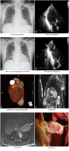
A 55-year-old man admitted with a 20-day history of intermittent chest pain. Electrocardiogram showed small Q wave and T wave inversion in the inferolateral leads. There were no specific findings on chest radiography (Fig. 1A). Echocardiogram showed akinesia and mild aneurysmal change of the left ventricular (LV) posterolateral wall (Fig. 1B). Coronary angiography demonstrated total occlusion of the mid left circumflex artery. Under the diagnosis of recent myocardial infarction, we performed aspiration thrombectomy and deployed two drug-eluting stents.
After an event-free 14-month follow-up period, we detected abnormal contour on repeat chest radiography (Fig. 1C). Echocardiography confirmed large out-pouching aneurysmal change of LV posterolateral wall with narrow neck containing hyperechoic thrombi (Fig. 1D).1-3) Multi-detector computed tomography revealed definite bulging-out contour of the LV wall (Fig. 1E).3) A fast, steady-state sequence of cardiac magnetic resonance imaging showed discontinuity of the myocardium and a bulging sac. The maximum width of the orifice was shorter than the maximum parallel internal diameter (Fig. 1F).4)5) On inversion-recovery-prepared breath-hold cine gradient-echo sequence, markedly delayed enhancement of the pericardium that forms the wall (white arrow), as well as linear hypointense thrombus were detected (Fig. 1G), which have been suggested as helpful additional features for differentiation between true and false LV aneurysms.4) Because of rupture risk, the patient was treated with LV aneurysmectomy. Following thoracotomy, we observed narrow-necked channel with a larger aneurysmal sac, which contains blood and thrombus (Fig. 1H). Histologic finding revealed fibrous pericar-dial tissue and thrombi with no myocardial elements. The patient remained complication free following the operation.
 XML Download