

 PDF
PDF ePub
ePub Citation
 Print
Print


Introduction
Kawasaki disease (KD) is an acute febrile exanthematous illness involving development of vasculitis in various tissue and organs, and frequently affects coronary arteries and cardiac tissue, causing coronary ectasia or aneurysm, myocarditis, valvulitis, and pericarditis.1) KD is now the most important cause of acquired heart disease in infants and children.2) These days, treatment of KD consists of intravenous immunoglobulin (IVIG) and oral aspirin, and IVIG is usually administered as a single infusion of 2 g/kg (high-dose IVIG).1) However, some clinical studies have shown that a single infusion of IVIG 1 g/kg (medium-dose IVIG) was also effective.3-7) Since the 1990s, Han et al.8) has administered medium-dose IVIG as the initial treatment for patients with KD, giving one more infusion after 48 hours if fever persisted, and results that such a mediumdose regimen have been good.
The cost of IVIG is high (about 40,000-45,000 won/2.5 g). For a patient weighing 20 kg and receiving high-dose IVIG, the cost of IVIG is about 640,000-720,000 won. If the patient receives medium-dose IVIG, the cost will be reduced to 320,000-360,000 won. Therefore, the authors think that the medium-dose regimen proffers an advantage over the high-dose regimen in view of cost-effectiveness, if the two regimens are equally effective. We therefore performed a retrospective clinical study to validate effectiveness of the medium-dose regimen in treatment of KD.
Subjects and Methods
Two hundred seventy four patients (162 males and 112 females; M:F=1.45:1) with KD were admitted to Bundang Jesaeng General Hospital from July 1998 to October 2007. The proportion of patients with KD was 1.57% of all patients admitted to the pediatric ward (with the exception of the neonatal unit) of the hospital during the study period. Diagnosis of KD was based on the presence of fever for 5 days or more and presentation with 4 or more of 5 principal clinical features (bilateral bulbar conjunctival injection; changes in lips, tongue, or oral mucosa; cervical lymphadenopathy greater than 1.5 cm in diameter; polymorphous skin rash; changes in extremities).9) In cases where a patient had fever for 7 days or more, and 3 or less of 5 principal clinical features, and other diseases were excluded, echocardiography was performed. If findings on echocardiography were abnormal {coronary arterial lesions (CAL), decreased ventricular systolic function, significant valvular regurgitation, and pericardial effusion}, the patient was diagnosed as having KD.1) CAL included ectasia and aneurysm. Normal internal diameters of coronary arteries were defined as 2.5 mm or less in infants less than 1 year old, 3 mm or less in children from 1 to 4 years old, and 4 mm or less in children 5 years or older. Significant valvular regurgitation was defined as mitral or aortic valvular regurgitation of grade 1 or more and tricuspid or pulmonary valvular regurgitation of grade 2 or more, according to the previous study.10)
Medium-dose IVIG (1 g/kg) was infused over 8-10 hours as the initial treatment as soon as the diagnosis of KD was made. If fever did not subside within 48 hours, one more infusion of medium-dose IVIG was given. If fever persisted or relapsed thereafter, one or more infusions of medium-dose IVIG was given again. Aspirin (30-50 mg/kg/day) was given orally in 3 divided doses from the time of diagnosis until one week after defervescence; the dosage was then reduced to 3-5 mg/kg/day in a single dose.
Patients treated with a single infusion of mediumdose IVIG were defined as group A, and patients treated with two or more infusions were defined as group B. Patient characteristics in both groups were compared; these included demographic features, duration of fever before and after treatment, laboratory findings, and rates of CAL. Continuous variables were described as mean±standard deviation. Statistical analyses were performed by means of the Student t-test for continuous variables and the Chi-square test for rates and proportions. P of less than 0.05 was considered statistically significant.
Results
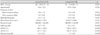
Among 274 patients, 220 of them showed improvement with a single infusion of medium-dose IVIG (group A; n=220; 80.3%). Forty one patients showed improvement with two infusions, 11 patients showed improvement with three infusions, and two patients showed improvement with four and five infusions each (group B; n=54; 19.7%). Male-to-female ratios (1.37:1 in group A vs. 1.84:1 in group B), age distributions (33.8±25.2 vs. 35.9±29.4 months), duration of fever before treatment (5.6±1.8 vs. 5.0±1.8 days), hemoglobin concentrations (11.2±1.0 vs. 11.4±0.8 g/dL), white blood cell counts (15,010±5,370 vs. 15,570±5,580/mcL), and platelet counts (374,000±126,200 vs. 358,500±138,100/mcL) did not differ significantly between the two groups (p>0.05) (Table 1).
Duration of fever after treatment was significantly longer in group B than in group A (0.5±0.8 days in group A vs. 4.5±2.6 days in group B), and concentrations of C-reactive protein (8.7±5.4 vs. 12.1±5.7 mg/dL), aspartate aminotransferase (69.2±110.8 vs. 150.4±245.7 U/L), alanine aminotransferase (82.7±111.2 vs. 156.3±202.4 U/L), and total bilirubin (0.5±0.7 vs. 1.1±1.4 mg/dL) were significantly higher in group B than in group A (p<0.005, respectively) (Table 1).
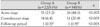
CAL were found in 51 patients (23.2%) in group A and 26 patients (48.1%) in group B who were in the acute stage (p<0.005). CAL were found in 14 patients (6.4%) in group A and 11 patients (20.4%) in group B who were in the convalescent stage (p<0.005). During the follow-up period, CAL disappeared in 13 of 14 patients in group A and in 7 of 11 patients in group B. A giant aneurysm of 8 mm or more in diameter was found in one patient in each group (0.5% in group A and 1.9% in group B; p<0.005) (Table 2). Three patients in group B were lost to follow-up.
Discussion
Suppression of acute inflammatory reactions and prevention or reduction of cardiac complications, such as coronary aneurysm are the goals of treatment in KD. Efficacy of IVIG in treatment of KD was first reported in 1984.11) IVIG was given at the dosage of 400 mg/kg/day for 3-5 consecutive days during the earlier period. It was reported years later that a single infusion of IVIG 2 g/kg suppressed acute symptoms of KD more rapidly, decreased duration of both intravenous injection and hospitalization, and effectively prevented coronary aneurysm.12-14) Thereafter, a single infusion of high-dose IVIG became the standard regimen in treatment of KD (the high-dose regimen).
Opinions have differed, however, on the effective dosage of IVIG in treatment of KD. Engle et al.3) reported that a single infusion of IVIG 1 g/kg was effective in treatment of KD, and Furusho et al.4) suggested that the dosage of IVIG should be 1 g/kg or more for effective prevention of CAL. Barron et al.5) compared effectiveness of a single infusion of IVIG 1 g/kg and infusions of 400 mg/kg/day for 4 days and reported that a single infusion of 1 g/kg suppressed fever more rapidly and prevented coronary aneurysm with equal effectiveness. Ihn et al.6) and Qin et al.7) compared effectiveness of IVIG 1 g/kg and 2 g/kg and reported that the two regimens were equally effective. Han et al.8) performed a clinical study to evaluate effectiveness of medium-dose IVIG during the 1990s. Results from that study showed improvement in 86% of patients with a single infusion of IVIG 1 g/kg and 14% showed improvement with two infusions of IVIG 1 g/kg. Rates of coronary aneurysm showed no significant difference from each other or from patients treated with a single infusion of IVIG 2 g/kg. Therefore, Han et al.8) thought that medium-dose IVIG was effective in the majority of patients with KD and that the mediumdose regimen was more advantageous than the highdose regimen in view of cost-effectiveness.
KD shows various degrees of severity and ranges from a mild illness that subsides quickly without complications to a very severe illness that is complicated by serious complications and can be fatal. Several scoring systems have been developed to identify patients at higher risk for coronary aneurysm so that they can be treated more aggressively. However, the scoring systems are not accurate, and their application in clinical practice is somewhat cumbersome. Therefore, the authors recommend the medium-dose regimen in which a single infusion of IVIG 1 g/kg is given as the initial treatment. A single infusion of medium-dose IVIG will be sufficient for the majority of patients suffering mildtomoderate KD, and one or more additional infusions can be given to the minority of patients who suffer severe KD and do not respond to a single infusion of medium-dose IVIG.
In this study, 80% of patients showed improvement with a single infusion of IVIG 1 g/kg (group A) and 20% showed improvement with two or more infusions of IVIG 1 g/kg (group B). These results were similar to those of the previous study by Han et al.8) Demographic features, duration of fever before treatment, hemoglobin concentrations, and white blood cell and platelet counts did not differ significantly between two groups. However, concentrations of C-reactive protein, aspartate aminotransferase, alanine aminotransferase, bilirubin, and rates of CAL were significantly higher in group B than in group A. It can be postulated that patients in group A suffered mild-to-moderate illness and patients in group B suffered severe illness. So the latter might require greater amounts of IVIG for suppression of more severe inflammatory reactions, and therefore develop CAL more frequently than the former. Therefore, the authors insist that even a single infusion of medium-dose IVIG is effective in the majority (about 80%) of patients, and one or more additional infusions are needed only in a minority (about 20%) of patients who do not respond to a single infusion of medium-dose IVIG. Use of the medium-dose regimen will reduce the cost of treatment by 40% roughly, compared with use of the high-dose regimen.
Some may have questions regarding the accuracy of diagnosis of KD in patients enrolled in this study. The authors made their diagnoses based on currently approved diagnostic criteria.1)9) The proportion of patients with KD was 1.57% of all patients admitted to the pediatric ward (with the exception of the neonatal unit) of Bundang Jesaeng General Hospital during the study period. This proportion was similar to the mean proportion of patients with KD (1.48%) in a recent nationwide survey.15) Therefore, the authors think that the diagnosis of KD in patients enrolled in this study was accurate.
In this study, the authors defined normal internal diameters of coronary arteries as 2.5 mm or less in infants less than 1 year old, 3 mm or less in children from 1 year to 4 years old, and 4 mm or less in children 5 years or older. The Japanese Ministry of Health defines normal internal diameters of coronary arteries as 3 mm or less in children 4 years or younger and 4 mm or less in children 5 years or older.16) In the opinion of the authors, however, the criteria of the Japanese Ministry of Health are too simple and may underestimate of rates CAL in small infants. Nakano et al.17) and de Zorzi et al.18) proposed that normal internal diameters of coronary arteries be 2.5 mm or less if body surface area (BSA) is less than 0.5 m2, 2.5-3 mm if BSA is 0.5-1 m2, and 3 mm or more if BSA is 1 m2 or more. Other criteria define normal internal diameters of coronary arteries as 2.5 mm or less in children weighing less than 12.5 kg, 2.5-3 mm in children weighing between 12.5 kg and 27.5 kg, and 3-5 mm in children weighing 27.5 kg or more.19) Therefore, the authors classified infants less than 1 year old separately, and, within this context, defined the normal internal diameter of coronary arteries as 2.5 mm or less. Rates of CAL in this study were higher than those in other studies. The reasons for these results could be explained by the following: The authors included mild coronary ectasia into CAL, and more infants may have been diagnosed with CAL by use of our detailed criteria on the normal diameter of coronary arteries in infants.
The limitations of this study are that it is a retrospective study, and we evaluated treatment outcomes in patients treated with medium-dose IVIG, instead of comparing them with those in patients treated with high-dose IVIG. However, in the previous study by Han et al.8) treatment outcomes in patients treated with medium-dose IVIG were similar to those in patients treated with high-dose IVIG.8) Therefore, the authors think that effectiveness of medium-dose IVIG is consistent and comparable with that of high-dose IVIG in the majority of patients with KD. In the minority of patients with severe KD, however, two or more infusions of medium-dose IVIG will be required. In conclusion, the authors think that the regimen with medium-dose IVIG (1 g/kg) is effective in the majority of patients with KD and proffers an advantage over the regimen with high-dose IVIG (2 g/kg) in view of cost-effectiveness.
 XML Download