

 PDF
PDF ePub
ePub Citation
 Print
Print


Introduction
Though percutaneous coronary intervention (PCI) is an effective treatment in the setting of coronary artery disease, contrast-induced nephropathy (CIN) is an important complication of iodinated contrast media infused during diagnostic or interventional procedures.1)2) CIN is the third leading cause of acute renal failure in admitted patients, accounting for 10% of all cases.3) This iatrogenic complication has been a concern to the interventional cardiologists in recent years because of its adverse effect on prognosis. The development of CIN is associated with an increased risk of death and late cardiovascular events after PCI such as myocardial infarction and target vessel revascularization.4-8)
CIN is commonly defined as a rise in serum creatinine of ≥25% or ≥0.5 mg/dL above the baseline value within 48 hours after contrast administration. On the basis of this definition, the overall incidence of CIN in the general population is reported to be 1.2 to 1.6%.9-11)
A key step in minimizing CIN is to identify patients at high risk. Known risk factors for CIN include chronic kidney disease (CKD), diabetes mellitus (DM), volume depletion, nephrotoxic drug therapies, hemodynamic instability, and intra-aortic balloon pump treatment.12-14) The additive nature of risk has allowed for the development of prognostic scoring systems, but the type and dose of contrast administration have not been fully analyzed in these scoring systems.15)16) Moreover, the American College of Cardiology/American Heart Association (ACC/AHA) guidelines for acute coronary syndromes in the setting of CKD list the use of isosmolal contrast media as a class I (Level A) recommendation.17) However, isosmolal contrast media is not administered for all patients in routine clinical practice, which creates discrepancies between the scoring systems and daily practice. To avoid such discrepancies, we investigated other possible risk factors for CIN in PCI patients who were administered an isosmolal contrast agent, iodixanol, as the only contrast media.
Subjects and Methods
In order to minimize the potential role of hemodynamic instability as a cause of postprocedural renal failure, patients were excluded who had ST-segment elevation myocardial infarction requiring primary PCI, or cardiogenic shock requiring treatment with an intraaortic balloon pump (IABP) or inotropic agents. Patients with serum creatinine >4.0 mg/dL, dependence on dialysis, neoplasms, vascular malformations, thrombocytopenia, or major bleeding (intracranial, intraocular, or retroperitoneal hemorrhage, clinical bleeding with hemoglobin drop >3.0 g/dL, or red cell transfusion ≥2 units) were also excluded. For patients who underwent more than one angiographic procedure during the study period, only the first PCI was included.
There were 956 patients who underwent PCI at Chonbuk National University Hospital between January 2005 and December 2006. Of these, we excluded 23 patients who had definite cause for renal dysfunction after PCI such as infection, renal artery thrombosis, hemolysis, rhabdomyolysis, renal stone, or prostate disease. We retrospectively analyzed the medical records of 537 patients who underwent PCI and were administered iodixanol (Visipaque®, GE Health Care, Cork, Ireland) as contrast media. The patients were divided into two groups: Group I (486 patients who did not develop CIN) and Group II (51 patients who developed CIN).
Elective PCI patients received routine hydration with 0.9% normal saline >1 mL/kg/hour from 4 to 12 hours before PCI and continuing after PCI. However, some patients requiring urgent PCI could not receive sufficient hydration {79 (16.3%) in Group I vs. 10 (19.6%) in Group II}. N-acetylcysteine 600 mg was administered twice on the day before the procedure and twice on the day after the procedure.
CIN was defined as a postprocedural increase in serum creatinine of ≥0.5 mg/dL or an increase of 25% from baseline. Absolute or relative increase in serum creatinine at 24 hours or 48 hours were compared to baseline serum creatinine, and CIN was diagnosed when alternative explanations for aggravation of renal dysfunction were ruled out. Per study design, blood samples were drawn before PCI and 24 hours after PCI to measure serum creatinine; further determinations of creatinine beyond 24 hours were done if clinically indicated.
Diagnostic angiography and PCI were performed after premedication with aspirin (at least 100 mg) and unfractionated or low molecular weight heparin. A loading dose of clopidogrel (between 300 mg and 600 mg) was administered before PCI. Coronary angiography was performed through the femoral or radial artery. Heparin was infused throughout the procedure to maintain an activated clotting time of 250 seconds or longer. Stents were deployed after prior balloon angioplasty, and administration of a platelet glycoprotein IIb/IIIa-receptor blocker was left to the decision of the surgeon. The appropriate length and diameter coronary stent was carefully selected to properly cover the target lesion. Each individual physician decided on selection of the transradial or transfemoral approach, contrast dose, interventional technique, supportive pharmacologic therapies, and post-dilatation with a non-compliant high-pressure balloon. Therapeutic decision-making and the need for interventional therapy in patients with unstable angina/non-ST-elevation myocardial infarction (NSTEMI) were left to the physician's choice according to the guidelines of the ACC/AHA/Society for Cardiovascular Angiography and Interventions (SCAI).18)19)
The baseline clinical characteristics, laboratory characteristics, and coronary angiography findings {including ACC/AHA classification, and Thrombolysis In Myocardial Infarction (TIMI) flow grade before and after PCI} were analyzed for groups I and II. To examine a report that each 3% periprocedural drop in hematocrit (known to correspond approximately to 1 g/dL of hemoglobin) resulted in development of CIN, we also analyzed the effect of a decrease in hemoglobin by ≤1 g/dL, 1-2 g/dL, ≥2 g/dL on the development of CIN in our study population.20)
For continuous variables, comparisons between the groups were done using Student's t-test. Fischer's exact test was used to evaluate the categorical variables. To test whether initial differences between the two groups influenced the differing results, multiple logistic regression analysis was performed after controlling the variables that were significantly different at baseline. All continuous variables are described as mean±standard deviation. All analyses were 2-tailed, with clinical significance defined as p<0.05. All statistical processing was done using SPSS-PC 15.0 (Statistical Package for the Social Sciences, SPSS-PC. Inc., Chicago, IL, USA).
Results
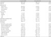
Baseline clinical and laboratory characteristics (Table 1) were not significantly different between the two groups. However, patients who developed CIN (Group II) had lower abdominal circumference, body weight, body mass index (BMI), baseline hemoglobin, and post-PCI hemoglobin levels.
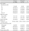
There was no significant difference between the two groups with regard to lesion characteristics by coronary angiography according to ACC/AHA classification or TIMI flow grade before PCI. Moreover, the drugs used for anticoagulation and the closure device were not different between the two groups. However, the TIMI flow grade after PCI was higher in patients who did not develop CIN (Group I) compared to the patients who developed CIN (Group II) (3.0±0.2 vs. 2.8±0.4, p=0.003) (Table 2). Indications for PCI were different between the two groups. The proportion of urgent PCIs (early invasive PCI rather than elective PCI) was higher in Group II (45.1%) than in Group I (35.2%) (p=0.009).
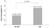
The proportion of patients who experienced a drop in hemoglobin of more than 1 g/dL was higher among patients who developed CIN (Group II, 54.9%) compared with patients who did not develop CIN (Group I, 39.3%) (p=0.036) (Fig. 1).
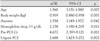
Multivariate logistic regression analysis was done both by statistically significant variables (body weight, baseline hemoglobin, post-PCI hemoglobin, pre-PCI Cr, post-PCI Cr, and TIMI flow grade after PCI) and conventional risk factors for CIN (age, anemia, and urgent PCI). Multivariate logistic regression analysis showed that the strong predictors for the development of CIN (Table 3) were consistent with previous studies; age, body weight, anemia, pre-PCI creatinine, and urgent PCI. Like other known risk factors for CIN, a decrease in hemoglobin by >1 g/dL was a strong predictor for the development of CIN {adjusted odds ratio (aOR)=2.238, p=0.012}.
Discussion
CIN is one of the most common causes of acute renal failure in hospitalized patients. The frequency of CIN has decreased over the past decade from a general incidence of -15% to -7%.15) CIN is associated with prolonged in-hospital stay and increased morbidity, mortality, and cost. In the Mayo Clinic registry, the inhospital death rate was 22% among the patients who developed CIN compared to only 1.4% in patients who did not develop CIN. Among hospital survivors with CIN, 1- and 5-year estimated mortality rates were 12.1% and 44.6%, respectively, much greater than the 3.7% and 14.5% mortality rates noted in patients without CIN (p<0.0001).1)
Although the pathogenesis of CIN is not completely known, multiple mechanisms may be involved. Generally, the pathophysiology of CIN assumes baseline reduced nephron number, with superimposed acute vasoconstriction caused by the release of adenosine, endothelin, and other renal vasoconstrictors triggered by iodinated contrast. After a very brief increase in renal blood flow, there is an overall -50% sustained reduction in renal blood flow lasting for several hours. Prolonged contrast transit time in the kidney increases the exposure of renal tubular cells to contrast. This stasis of contrast in the kidney allows for direct cellular injury and death in renal tubular cells. Furthermore, the sustained reduction in renal blood flow to the outer medulla leads to medullary hypoxia, ischemic injury, and death of renal tubular cells.21)
Many individual risk factors have been reported in relation to the development of CIN. The best recognized non-modifiable risk factors are older age, DM, preexistent renal insufficiency, congestive heart failure, hemodynamic instability, and nephritic syndrome. Modifiable risk factors include volume depletion, volume of contrast media, nephrotoxic drug use, low serum albumin level (<35 g/L), and anemia.22)23)
Dangas et al.24) showed that the baseline hematocrit level is an independent predictor of CIN in patients with CKD (OR=0.95, p<0.00001). Nikolsky et al.20) reported that lower baseline hematocrit was an independent predictor for CIN in 6,773 consecutive patients treated with PCI. Moreover, the CIN rate steadily increased with baseline hematocrit quintile decrements (from 10.3% in the highest quintile to 23.3% in the lowest quintile) (p<0.0001). Each 3% drop in hematocrit (known to correspond to approximately 1 g/dL of hemoglobin) resulted in a 30% increase in the risk of CIN (p<0.0001) in patients with baseline CKD and a 26% increase in the risk of CIN in patients without CKD (p<0.0001).
On multiple logistic regression analysis in the present study, traditional risk factors for CIN such as age, body weight, anemia, pre-PCI creatinine, and urgent PCI showed a significant association with CIN, consistent with previous studies. Interestingly, the present study indicates that a hemoglobin drop (>1 g/dL) during PCI is an important predicting factor for CIN, just like other known risk factors. Even though the association between hemoglobin drop and higher rates of CIN is not clearly understood, patients with a greater decrease in hemoglobin were older, had lower baseline hemoglobin, and required more urgent PCI.16)25) Periprocedural hemoglobin drop was likely related to blood loss incurred through puncture site hematoma formation, and through catheter aspiration. Those had an additive impact to the prevention of CIN in real clinical practice.
In the present study, all patients were administered the lowest nephrotoxic isosmolal contrast agent and there was no statistical difference in administered contrast agent volume. The clinical meaning of the present study is that physician should make every effort to decrease periprocedural bleeding, even in a setting of lowest nephrotoxic contrast agent and N-acetylcysteine prevention.
However, our small, single tertiary center, retrospective study has several limitations. First, we may have underestimated the incidence of CIN because we did not check routine serum creatinine at 72 hours after PCI. Moreover, routine hydration could not be carried out in some patients who required urgent PCI within 4 hours. These discrepancies should be considered in future investigations. Second, our study did not address the other important variables related to CIN, such as hydration volume, urine output, hemodynamic parameters, procedure time and nephrotoxic medications. This is major limitation of our study. Third, our study was directed at low risk patients. We excluded patients who were at high risk for CIN, such as those with serum creatinine >4.0 g/dL, primary PCI, cardiogenic shock and major bleeding. Fourth, we did not analyze all red blood cell data on erythropoietin levels, plasma volume, and red blood cell mass that would provide more information on the development of CIN.
In summary, a periprocedural drop in hemoglobin of more than 1 g/dL is another important independent predictor for CIN, even in patients administered the lowest nephrotoxic contrast agent, iodixanol, during PCI. Because even a small amount of procedure-related blood loss is associated with the development of CIN and leads to poorer clinical outcomes, interventional cardiologists should maker every effort to reduce blood loss during PCI.
 XML Download