

 PDF
PDF ePub
ePub Citation
 Print
Print


Introduction
In 2020, 300 million adults worldwide are expected to suffer from diabetes and the majority will be type 2 diabetes.1) The incidence of type 2 diabetes has increased rapidly, especially in Asian countries, and cardiovascular events are the most important causes of death in type 2 diabetic patients.2) Despite great advances in coronary artery intervention, reduction in cardiovascular events and mortality in patients with type 2 diabetes has not been remarkable.3)
Thiazolidinediones (TZDs), including pioglitazone and rosiglitazone, were introduced as insulin sensitizers. Pioglitazone and rosiglitazone are peroxisome proliferator-activated receptor (PPAR)-γ agonists, have been known for their anti-inflammatory effects independent of blood glucose control.4)5) Because chronic low-grade inflammation results in atherosclerosis and cardiovascular diseases, TZDs were expected to have positive effects on atherosclerosis progression. Rosiglitazone was shown in a meta-analyses to increase the risk of ischemic events, including myocardial infarction.6) On the other hand, two recent studies revealed that carotid intima media thickness regressed in patients treated with pioglitazone, compared to patients treated with glimepiride while both agents demonstrated similar HbA1c lowering effects.7)8) The Pioglitazone Effect on Regression of Intravascular Sonographic Coronary Obstruction Prospective Evaluation (PERISCOPE) trial showed that pioglitazone slowed the progression of atherosclerosis compared to glimepiride in patients with type 2 diabetes.9) Pioglitazone compared with rosiglitazone appears to have more anti-atherogenic effects independent of the glucose lowering effects. The purpose of our study was to determine the effects of pioglitazone, when compared with placebo, on atherosclerotic progression and neointima formation by intravascular ultrasonography (IVUS) in type 2 diabetic patients following zotarolimus-eluting stent (ZES) implantation. We also investigated the changes in the levels of inflammatory and insulin resistance markers, such as homeostatic model assessment (HOMA)-index and retinol binding protein (RBP)-4, which could be affected by pioglitazone.
Subjects and Methods
Study population
Patients were eligible for this study if they were 40 to 75 years of age and had type 2 diabetes. A total of 37 patients with high-grade coronary artery lesions, defined as stenosis above 70% of lumen diameter, were prospectively enrolled following ZES implantation at the Korea University Anam Hospital cardiovascular centers from March 2007 to January 2008. Eligible patients (n=37, 15 women and 22 men) were randomly assigned to receive either pioglitazone 15 mg (19 patients) or placebo (18 patients), in addition to standard diabetic management. We excluded patients with acute myocardial infarction, left main coronary lesion, prior history of interventional or surgical treatment for coronary artery disease, heart failure defined as ejection fraction less than 45%, hepatic dysfunction, defined as aspartate aminotransferase or alanine aminotransferase more than twice the upper limit, cerebrovascular disease, uncontrolled arrhythmia within 3 months, serum creatinine greater than 2.0 mg/dL, expected life expectancy of less than 1 year and previous use of PPAR-γ agonists within 3 months before the enrollment.
Study design
This was a prospective, randomized, single-blinded, study with an 8-month follow-up period, to evaluate the effects of pioglitazone in reducing atherosclerosis progression in type 2 diabetic patients. Most patients in both groups were taking anti-diabetic medication or insulin (85.7% vs. 78.5%) at enrollment. Aspirin 100 mg/day and clopidogrel 75 mg/day were maintained in all participants. Balloon angioplasty and ZES implantation were performed based on standard clinical practice. This study was approved by the institute review board, and all participants gave written informed consent at study entry. Eligible patients were randomly assigned to the pioglitazone group (n=19) and the placebo group (n=18), and then underwent IVUS and blood sampling. All other medications were continued, and all patients showed good compliance. After 8 months, clinical evaluation, blood sampling and repeat IVUS were performed to determine atherosclerosis progression. The results were analyzed by the intent-to-treat principle. Major adverse cardiovascular events (MACE), such as death, non-fatal myocardial infarction and the need for repeated target vessel or target lesion revascularization were noted at the 8-month follow-up.
Angiographic analysis and intravascular ultrasound measurements
Coronary angiograms were obtained at baseline, immediately after stenting and after 8 months. Two identical orthogonal views were obtained in each patient after intracoronary administration of nitrates and stored on digital CD-ROM. All angiographic and clinical data were analyzed in the Korea University Anam Hospital core laboratory. End-diastolic frames were chosen for quantitative analysis, which was performed using a computer-based TCS system, Version 2.02 (Medcon Inc., Tel-Aviv, Israel) by an operator who was blinded to the patient's information. The reference diameter, minimal luminal diameter, percentage of stenosis and lesion length were calculated as the average value of 2 orthogonal views. The same views and calibration were used at follow-up angiography. The average diameter of normal segments proximal and distal to the treated lesion was defined as the reference diameter. Re-stenosis was defined as stenosis of greater than 50% of the luminal diameter. Balloon angioplasty and ZES implantation were performed according to standard clinical practice. IVUS examination was performed after ZES implantation and at the 8-month follow up. The ultrasound probe was inserted and went along the guide wire into the distal part of the culprit vessel, and a motor drive was connected and withdrew the probe at a rate of 0.5 mm/s. A follow-up IVUS study was done the same way on the same lesions. We examined ath-erosclerosis progression 20 mm distal and proximal to the ZES during the 8-month follow-up, and neointima formation was observed at the stented sites at 8 months. Digitized images in 1.0 mm intervals were analyzed by an operator blinded to clinical information. Vessel area, defined as the internal volume of the external elastic membrane and the lumen area were measured at each cross-section. All measured vessel areas at each cross-section were added and then divided by the number of cross-sections in order to obtain the average vessel volume per unit area. The average lumen volume per unit area was calculated the same way. Atheroma volume was calculated by subtracting lumen volume from vessel volume, and atherosclerosis progression was calculated by subtracting atheroma volume at the beginning of the study from the atheroma volume at the 8-month follow-up. Neointima volume was calculated by subtracting the lumen volume from the stent volume at the 8-month follow-up.
Laboratory analysis
Inflammatory markers, such as tumor necrosis factor (TNF)-α, interleukin (IL)-6 and high-sensitivity C-reactive protein (hs-CRP) and insulin resistance markers such as basal insulin, HOMA index and RBP-4 were measured in both groups at the beginning of the study and at the 8-month follow-up visit. Venous blood samples were drawn from each patient after 8 hours or overnight fasting. Blood samples were centrifuged to obtain plasma, which in turn was stored at -80℃. TNF-α was measured by a sandwich enzyme linked immuno sorbent assay (ELISA), with a minimum detectable level of 0.5 pg/mL (ALPCO Diagnostics, Salem, NH, USA). Undetectable TNF-α values were recorded as 0.4 pg/mL. High-sensitivity IL-6 was also measured by a sandwich ELISA, with a minimum detectable level of 0.16 pg/mL (ALPCO Diagnostics, Salem, NH, USA). hsCRP concentration was quantified using a latex nephelometer II (Dade Behring Inc., Newark, DE, USA). Serum insulin was measured by microparticle enzyme immunoassay. Insulin resistance was assessed by the HOMA, calculated as {fasting serum insulin (µU/mL)×fasting serum glucose (mmol/L)}/22.5. Plasma RBP-4 concentration was assessed by ELISA. Total cholesterol, triglyceride, high density lipoprotein-cholesterol and low density lipoprotein-cholesterol concentrations were determined by the enzymatic methods using standard biochemical procedures on a B.M. Hitachi automated clinical chemistry analyzer (Hitachi, Tokyo, Japan).
Statistical analysis
Data are expressed as mean±SD for continuous variables, and data for the categorical variables are expressed as the number and the percentage of patients. Fisher's exact test or the chi-square test was used for categoric variables. The results between groups were compared by the Mann-Whitney U test, and comparisons between results pre and post treatment were analyzed by the Wilcoxon matched-pairs single-rank test. Simple correlation analysis was performed by the Spearman's rank order correlation. A p less than 0.05 was considered statistically significant. Statistical Package for the Social Sciences (SPSS) software (version 10.0) was used for analyses (SPSS Inc., Chicago, IL, USA).
Results
Patient population and clinical findings
Risk factors such as hypertension and smoking were the same between the two groups (Table 1). Target vessels were similar between the two groups. Baseline reference diameter, minimal lumen diameter and stenosis diameter were similar between the two groups (Table 1). Most patients were taking an oral diabetic medication before enrollment (89.5% vs. 77.8%) (Table 1). The use of angiotensin converting enzyme inhibitors or angiotensin receptor blockers were similar between the two groups, and atorvastatin 10 mg was maintained in all participants unless contraindicated.
There were no significant differences in baseline angiographic characteristics between the two groups, and the 8-month follow-up minimal lumen diameters were higher in the pioglitazone group, albeit without statistically significant difference (Table 2).
Changes in atherosclerosis progression
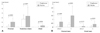
Baseline vessel volume, lumen volume and atheroma volume were similar between the two groups in atherosclerosis progression 20 mm proximal and distal to the stent (p=not significant). Mean change in atheroma volume was significantly lower in the pioglitazone group, compared to the placebo group at 8-month follow-up (0.06±0.73 vs. 1.16±1.41 mm3/mm, p=0.024, respectively) (Table 3). Atherosclerosis progression at 8 months was significantly greater compared to that of baseline in the placebo group (7.77±4.47 vs. 6.61±4.66 mm3/mm, p=0.016, respectively). On the other hand, there was no significant atherosclerosis progression at 8 months compared to baseline in the pioglitazone Group (5.24±2.89 vs. 5.18±2.69 mm3/mm, p=0.826). Changes in atheroma volume 20 mm proximal and distal to the stent were significantly lower in the pioglitazone group (Fig. 1A). Changes in atheroma volume are further divided into segments 5 mm immediately proximal and distal to the stent, and 5 to 20 mm proximal and distal to the stent. Changes in atheroma volume 5 mm immediately proximal and distal to the stent were higher than that of atheroma volume 5 to 20 mm proximal and distal to the stent (Fig. 1B).
In-stent neointima volume changes during follow-up
The pioglitazone group showed significantly lower neointima volume compared to the placebo group (1.74±0.93 vs. 2.42±1.98, p=0.007, respectively) (Fig. 1A). Two cases of instent re-stenosis were noted in the placebo group, and all of them belong to the diffuse proliferative type. One case of instent re-stenosis in the pioglitazone group belong to the focal type. No correlation was found between change in atheroma volume and neointima volume in all 37 patients (r=0.126, p=0.521).
Changes in the levels of inflammatory and insulin resistance markers
Baseline IL-6 and TNF-α concentrations were similar at baseline between the two groups. However, IL-6 and TNF-α concentrations at 8 months were significantly lower in the pioglitazone group compared to the placebo group (Table 4). Although fasting basal insulin and HOMA-I were similar at baseline between the two groups, 8-month follow-up fasting basal insulin and HOMA-I were significantly lower in the pioglitazone group, compared to the placebo group (Table 4). Moreover, adiponectin levels increased significantly only in the pioglitazone group. Fasting glucose, RBP-4, and hsCRP concentrations showed no significant difference between the two groups during the 8-month follow-up period. Moreover, changes from baseline in lipid profiles showed no significant difference between the two groups (Table 4). Changes from baseline in IL-6 (r=0.354, p=0.032) and TNF-alpha (r=0.412, p=0.043) levels demonstrated positive correlations with neointima volume in the pioglitazone group. The rates of MACE during the follow-up period were similar between the 2 groups.
Discussion
Retinopathy, neuropathy and nephropathy are the major microvascular complications in diabetes, and many studies revealed that intensive glycemic control reduced these microvascular complications.10) Oral glucose lowering medications, such as glimepiride and metformin, were not effective in lowering the incidence of macrovascular complications present as cardiovascular disease in type 2 diabetic patients.11) PPAR-γ agonists, such as pioglitazone and rosiglitazone are known insulin sensitizers. They demonstrate anti-inflammatory and anti-proliferative effects in addition to glycemic control.5) Although the exact mechanisms on how TZDs improve insulin sensitivity are not completely revealed, TZDs may improve insulin sensitivity by stimulating adipose differentiation and regulating genes that encode GLUT4.12)13) This insulin sensitizing effect is considered to be good influence on cardiovascular diseases, because insulin resistance is considered non-traditional risk factors for cardiovascular disease in diabetic patients.14) Previous data suggested that TZDs could have anti-atherogenic effects, and pioglitazone slowed atherosclerosis progression in the carotid artery.8) The PERISCOPE study revealed the effects of pioglitazone on lowering the progression rate of coronary atheroma, compared to glimepiride,9) and the effect of slowing atherosclerosis progression was not associated with the glucose lowering effect of pioglitazone.9) The relationship between atherosclerosis progression and improvement in insulin resistance was not clearly revealed in our study, probably due to the small study population. It must be emphasized that the majority of patients in the PERISCOPE study were Caucasians, who showed a high BMI (>32 kg/m2 in both glimepiride and pioglitazone groups) and high fasting serum insulin levels (>21 µIU/mL in both glimepiride and pioglitazone groups) at the beginning of the study. Most patients in the PERISCOPE study had type 2 diabetes with high insulin resistance, which constituted the major subgroup of type 2 diabetes in Western countries. However, Korean type 2 diabetic patients are known to differ from that of Western countries: Korean type 2 diabetes have relatively lower BMI with low fasting serum insulin levels.15-17) Most patients in our study had insulin secretory defects.15-17) Our study population also showed relatively low BMI (average 25.5 kg/m2) and low fasting serum insulin level (average 11.4 µIU/mL), compared to that of the PERISCOPE study. One might ask whether pioglitazone could have a similar effect of reducing the rate of atherosclerosis progression in Koreans, as demonstrated in a previous trial.9)
We also compared changes in the levels of RBP-4 in addition to HOMA index as markers of the insulin sensitivity, to determine in detailed the relationship between reduction in atherosclerosis progression and reduction in insulin resistance. However, no relationship between atherosclerosis progression and insulin resistance was found. Furthermore, we could not determine any relationship between RBP-4 concentrations with atherosclerosis progression and neointima formation, and there was no correlation between atherosclerosis progression and changes in HOMA-index levels. IL-6 and TNF-α levels at 8-month follow-up decreased significantly in the pioglitazone group, compared to the placebo group. IL-6 and TNF-α are representative of adipokine. Many studies showed the effects of pioglitazone on reducing inflammatory markers, such as TNF-α levels,18) and some animal studies showed neutralization of TNF-α caused significant increase in insulin sensitivity by increasing peripheral uptake of glucose.19) We also showed that pioglitazone has IL-6 and TNF-α lowering effects, thereby reducing atherosclerosis progression.
No correlation between atherosclerosis progression and neointima volume was found in our study. Several hypotheses could be put forward about this finding. First, a false negative result may be due to the small population size. Second, there could be a different mechanism between atherosclerosis progression and neointima proliferation. Third, zotarolimus compared may have stronger effects on neointima proliferation, and zotarolimus had a small effect on atherosclerosis progression 20 mm proximal and distal to the stent. One animal study showed TZDs decreased neointima proliferation by decreasing smooth muscle cell growth and inflammation.20) Our study did not address the mechanisms of pioglitazone in reducing atherosclerosis progression and neointima proliferation. The PROactive study showed MACEs reduction in the pioglitazone groups, compared to the placebo group.21) Decrease in atherosclerosis progression and neointima proliferation in the pioglitazone group did not translate into benefits in MACEs at the 8-month follow-up in our study, and a long-term study with larger study population is warranted. We did not find any complications with pioglitazone therapy, such as edema, weight gain and bone fracture during the 8-month follow-up period.
In conclusion, pioglitazone treatment, when compared with placebo, was associated with significant reduction in atherosclerosis progression and neointima proliferation in patients with type 2 diabetic following ZES implantation, and these findings were associated with the anti-inflammatory effects of pioglitazone.

 XML Download