

 PDF
PDF ePub
ePub Citation
 Print
Print


Introduction
The worldwide prevalence of diabetes mellitus (DM) has been reported to be higher than 170,000,000 patients, and it is expected to increase to more than 360,000,000 patients by 2030.1) Among patients with coronary artery diseases (CAD), the prevalence of DM has been reported to be even higher, and, conversely, the prevalence of cardiovascular diseases (CVD) is relatively higher in patients with DM.2)3) In the Cardiovascular Health Study, which is a population based study in adults aged 65 years or older, Barziay et al.4) reported that the prevalence of clinical or subclinical CVD was 70% in those with normal glucose, 77% in those with an impaired fasting glucose and 86% in those with a newly diagnosed or known DM. As blood glucose levels increase, the incidence of clinical or preclinical atherosclerosis also increases.4)
Insulin resistance (IR) has been proposed to be a risk factor for CVD, together with hyperglycemia, dyslipidemia, hypertension, etc.5) IR can be measured with a homeostasis model assessment-IR (HOMA-IR)6) assay and insulin sensitivity can be measured with a quantitative insulin-sensitivity check index (QUICKI).7) The Verona Diabetes Complication study, which was conducted in patients with type 2 DM, showed that HOMA-IR was correlated with the prevalence of CVD.5) Some studies suggested that IR can be defined as a HOMA-IR score of ≥3.0-3.5 in Korean patients, and showed that as HOMA-IR was increased, risk factors for metabolic syndrome were also increased.8-10) However, Adler et al.11) reported that IR and insulin sensitivity were associated with risk factors of CVD including age, body mass index (BMI), triglycerides, cholesterol, and systolic blood pressure, but not with DM, an independent predictor of CVD in patients.
On the other hand, carotid intima-media thickness (CIMT) and vascular endothelial function, which have frequently been used as a surrogate marker of atherosclerosis, have also been reported to be independent risk factors for CVD. These surrogates are associated with risk factors of CVD including hypertension, smoking, DM and dyslipidemia.12-15)
Therefore, we sought to determine, in patients with coronary atherosclerosis, the prevalence of impaired glucose tolerance (IGT) and DM, and IR and insulin sensitivity. The second purpose of this study was to assess the relationship between (a) CIMT and (b) endothelial function, IR and insulin sensitivity in this study population.
Subjects and Methods
Subjects
Study subjects consisted of 187 consecutive patients who underwent coronary angiography (CAG) at Konyang University Hospital from September 2003 to May 2004.
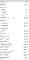
CAD was defined by stenosis ≥50% of the diameter of the lumen of a major epicardial coronary artery (left anterior descending artery, left circumflex artery or right coronary artery). Based on this definition, 88 patients (47.9%) had CAD and 99 patients (52.9%) had mild stenosis (<50%) (Table 1). Coronary risk factors including hypertension and smoking and clinical parameters including blood pressure, height and body weight were evaluated in all patients. We also measured cholesterol, triglyceride, low-density lipoprotein-cholesterol (LDL-C), high-density lipoprotein-cholesterol (HDL-C), high sensitivity C-reactive protein (hs-CRP), uric acid and homocysteine.
Oral glucose tolerance test, insulin resistance and insulin sensitivity
For the oral glucose tolerance test (OGTT), fasting plasma glucose, insulin and the concentration of C-peptide were measured after the patient had been at least 8 hours in a fasting state. Plasma glucose level was measured again at 60 and 120 minutes after intake of 75 g of glucose. Based on the results of the OGTT, new DM was defined as a fasting blood sugar (FBS) level ≥126 mg/dL or a plasma glucose level (120 minutes) ≥200 mg/dL. IGT was defined at 120 minutes as a plasma glucose level >140 mg/dL but <200 mg/dL. Patients who were receiving treatment for underlying DM were defined as "known DM".
Calculation of IR was done based on the concentrations of FBS and insulin. HOMA-IR was calculated based on the formula reported by Matthews et al.6) as shown below:
HOMA-IR={Fasting serum insulin value (in mU/L)×Fasting plasma glucose (in mg/dL)}/405
For the measurement of insulin sensitivity, the QUICKI index was calculated based on the methods reported by Katz et al.7) as shown below:
QUICKI=1/[{log(fasting insulin in mU/l)+log (fasting plasma glucose in mg/dL)}]
Coronary angiography
CAG was performed using a standard technique through the right femoral or radial artery. CAG was analyzed using online-quantitative coronary angiographic analysis (Philips, Integris c5000, Amsterdam, Netherland). Significant stenosis was defined as ≥50% diameter stenosis.
Measurement of endothelial function and carotid intima-media thickness
Flow-mediated brachial artery dilatation (FMD) was measured using high-resolution ultrasound (Hewlett-Packard Sonos 5500, Palo Alto, CA, USA) with an 11-3L (3-11 MHz) probe for evaluation of endothelial function. FMD was measured using methods that were proposed by Celermajer et al.16) in 1992. At first, the brachial artery was defined by the optimal gain control of the high resolutionultrasound three to five centimeters above the elbow. Then, the media to media distance of the brachial artery was measured at end diastole and the time velocity integral was obtained in the center of the brachial artery by Doppler ultrasound. The site at which the probe was positioned was marked and the landmark of the ultrasound image including branch and plaque was used for measurement of the same brachial artery site during the hyperemic phase. After measuring the brachial artery diameter and time velocity integral, the cuff of the sphygmomanometer was inflated up to 60 mmHg after the flow of the brachial artery had disappeared from the forearm for 5 minutes. Hyperemic blood flow was induced after decompression of the blood pressure cuff. The media-to-media distance and the time velocity integral at the same brachial artery site was measured 1 minute after decompression of cuff. The FMD was expressed as % change in the diameter of the hyperemic brachial artery relative to the baseline brachial artery diameter.
CIMT was also measured using high-resolution ultrasonography with the same product specifications, during which patients were placed in the supine position with their head extended into a dark room where the temperature and humidity were adjusted and the left common carotid artery was measured. The CIMT was measured in a 10 mm long segment just proximal to the carotid bulb in the common carotid artery and defined as the distance from the leading edge of the lumen-intima interface to the leading edge of the media-adventitia interface of the far wall. M'ATH® software (version 2.01, METRIS Co., Argenteuil, France) was used to determine accuracy and repeatability. M'ATH® software measures CIMT using a semi-automatic method. This digitalized measurement method has been reported to be greater than 4 fold more accurate than the conventional type of passive-mode measurement.17) To obtain optimal images, the depth and gain of ultrasonography were controlled. Depth control of the ultrasonography was fixed in a consistent manner for the purpose of calibration. An ultrasonographic gain was controlled to obtain images indicating the optimal intima, media and adventitia during the test. After optimal images were obtained, the imaging data were stored in a computer in which M'ATH® software was installed. M'ATH® software automatically calculates the mean for IMT. Testers selected the areas where the images were clearly visible at a 1 cm proximal location from the carotid bulb or they selected the interval whose length was approximately 1 cm with a quality index ≥0.60 to measured the IMT using M'ATH® software.
Statistical analysis
A comparison of data was made using the Statistical Package for the Social Sciences (SPSS) statistics program (version 12.0, USA). Then, the characteristics of patients were expressed as mean±SD. An analysis of the effects of each risk factor on atherosclerosis indicators was done using Pearson correlation analysis. Correlations between the tertiles of HOMA-IR/QUICKI and CIMT and endothelium-dependent vasodilatory responses were analyzed using ANOVA. A p of <0.05 was considered statistically significant.
Results
Clinical characteristics
Study subjects consisted of 187 patients {mean: 61.3±9.6 year-old, male gender 50.3% (n=94)} (Table 1). The prevalence of hypertension, DM, dyslipidemia and smoking was, respectively, 46% (n=86), 56.7% (n=106), 29.4% (n=55) and 25.7% (n=48). CAG showed that 88 patients (47.1%) had significant CAD. In these patients, the prevalence of single vessel disease was 24.1% (n=45), two vessel disease 15% (n=28) and three vessel disease 8% (n=15). The reason that CAG was done included stable angina in 166 patients (88.8%) and acute coronary syndrome (ACS) in 21 patients (11.2%). Mean BMI was 24.5±3.0 kg/m2 and left ventricular ejection fraction measured by transthoracic echocardiography was 64.7±10.3%. Laboratory tests showed a total cholesterol of 176.3±42.0 mg/dL, a triglyceride level of 155.5±83.2 mg/dL, an LDL-C of 108.0±34.1 mg/dL, an HDL-C of 40.1±9.9 mg/dL, an FBS of 117.1±39.6 mg/dL, homocysteine levels of 10.3±4.2 mg/dL, hs-CRP of 0.284±0.32 mg/dL and a uric acid of 5.0±1.6 mg/dL. Mean CIMT was 0.9±0.2 mm and mean FMD was 4.8±2.0%.
The prevalence of diabetes mellitus
Among all patients, DM was already known in 33.7% (n=63), new DM was found in 23% (n=43) and IGT was found in 31% (n=58). Of patients who already had DM, 16 were receiving insulin therapy and 47 were receiving oral anti-hyperglycemic agents. FBS and fasting insulin levels were 93.2±8.2 mg/dL and 8.9±2.1 IU/L, respectively, in non-DM patients, were 101.3±11.3 mg/dL and 10.2±4.1 IU/L in IGT patients, were 123.3±47.4 mg/dL and 11.1±7.3 IU/L in new DM patients, and were 138.2±40.1 mg/dL and 10.3±5.4 IU/L in known DM patients. The prevalence of DM did not have any difference between stable angina group and ACS group (46.3% vs. 46.1%) (p=ns). The prevalence of DM was 43.5% (n=22) in patients with single vessel disease, 58.2% (n=25) in patients with multi-vessel disease and 43.5% (n=42) in patients without significant stenosis. The prevalence of DM was increased in patients with multi-vessel disease, but this change was not statistically significant.
Correlations between insulin resistance, insulin sensitivity and indicators of atherosclerosis
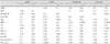
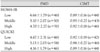
Mean HOMA-IR was 2.97±1.8 and mean QUICKI was 0.58±0.1. Using the definition of IR being HOMA-IR ≥3.0, there were 71 patients (38%) who had IR. The prevalence of IR was 21% (n=5) in non-DM patients, 34% (n=20) in patients with IGT, 44% (n=19) in patients with new DM and 42% (n=27) in patients with known DM (Table 2). The prevalence of IR was 35% (n=16) in patients with single vessel disease, 43% (n=18) in patients with multi-vessel disease and was 37% (n=37) in patients without significant stenosis. The prevalence of IR was increased in patients with multi-vessel disease, but this change was not statistically significant. There were significant positive correlations between HOMA-IR and BMI (r=0.28, p=0.0001) and HOMA-IR and triglyceride (r=0.2, p=0.01). But there were no correlations between HOMA-IR and FMD (r=-0.23, p=0.41) or CIMT (r=0.10, p=0.23). There were significant negative correlations between QUICKI and BMI (r=-0.26, p=0.0004), total cholesterol (r=-0.15, p=0.04), triglycerides (r=-0.21, p=0.004) and LDL-C (r=-0.17, p=0.02). But there were no correlations between QUCKI and FMD (r=-0.07, p=0.41) or CIMT (r=-0.13, p=0.13) (Table 3). Study subjects were divided into three groups according to HOMA-IR and QUICKI. CIMT and endothelium-dependent vasodilatory responses did not show any differences between groups (Table 4).
Correlations between Endothelium-Dependent Vasodilatation, intima-media thickness and risk factors
Among all patients, an analysis of the correlations between endothelium-dependent vasodilatation, CIMT and cardiovascular risk factors were done. CIMT was correlated with total cholesterol (r=0.19, p=0.02) and LDL-C (r=0.26, p=0.002). Endothelium-dependent vasodilatation was correlated with age (r=-0.19, p=0.02) and homocysteine level (r=-0.23, p=0.01). However, CIMT and endothelium-dependent vasodilatation did not correlate with HDL-C, triglyceride, uric acid or FBS (Table 3).
Discussion
It has been reported that the worldwide prevalence of DM is rising. In Korea, according to reports by Kim et al.18) in 2005, the prevalence of DM was 2.26% in adult males and 2.03% in females and was persistently increasing.
Major outcomes of the current study are that the prevalence of known DM (33.7%, n=63), new DM (22%, n=43) and IGT (31%, n=58) are high in patients with coronary atherosclerosis. According to other studies which were done in CAD patients, however, the prevalence of DM at the time of admission was 27%.19) In the current study, the prevalence of DM was 55.7% (known DM 33.7% and new DM 22%). A single measurement of FBS in patient with ACS may have contributed to the high prevalence of DM in our study. And, the prevalence of DM can vary depending on study subjects. Therefore, our study does not represent the true prevalence of DM in Korean patients with CAD. However, OGTT was done for all subjects to evaluate DM and IGT, and DM was newly diagnosed in many patients. Accordingly, more efforts are needed to evaluate DM in patients with coronary atherosclerosis. The prevalence of DM and IR can vary according to the number of affected blood vessels. In the current study, however, the prevalence of DM and IR was increased, but the increases were not statistically significant. The number of enrolled patients was relatively small. As mentioned earlier, the prevalence of DM and IR were assumed to increase due to the effects of other extrinsic factors.
In a study of Koreans, IR was defined as HOMA-IR ≥3.0-3.5.8-10) Also in the current study, cases of HOMA-IR ≥3.0 were defined as IR. In patients with coronary atherosclerosis, which included both those with DM and non-diabetic patients, the number of patients with IR was 71 (38%), and this corresponded to a relatively greater figure.
Ohnishi et al.20) reported that IR correlated with coronary risk factors including blood pressure, BMI, triglycerides and LDL-C. In the current study, IR measured by HOMA-IR positively correlated with BMI and triglycerides and insulin sensitivity and QUICKI scores negatively correlated with BMI, total cholesterol and LDL-C. CIMT and endothelium-dependent vasodilatation are used as surrogate marker of atherosclerosis and associated with coronary risk factors. CIMT was associated with age, sex, cholesterol, LDL-C and pulse pressure.21) Endothelium-dependent vasodilatation was associated with age, sex and the number of risk factors of atherosclerosis.22) Also in the current study, these indicators were associated with coronary risk factors. But there were no significant correlations between IR, insulin sensitivity, endothelium-dependent vasodilatation and CIMT. According to the report by Giannini et al.23) in obese children at the prepubertal period, as HOMA-IR was increased CIMT was also increased. Makino et al.24) reported that there was a significant correlation between endothelium-dependent vasodilatation and IR in patients with DM. The current study was conducted in patients who had coronary atherosclerosis, which showed that CIMT was 0.91±0.2 mm on average. According to the report by Bae et al.21) mean CIMT was 0.63±0.11 mm in Korean people. In the current study, however, CIMT was increased, and this may explain the discrepancy from other studies.
The current study has some limitations. This is not a case controlled study and the number of study subjects was relatively small. Also, we mostly studied cases of advanced coronary atherosclerosis. There were no clinical follow-up data for patients who were newly diagnosed with DM or IGT. Further studies are now needed to evaluate the prognostic value of active diagnosis of in patients with coronary atherosclerosis.
In conclusion, this study showed that the prevalence of new DM and IGT is relatively higher in older patients with coronary atherosclerosis than in older individuals in the general population. Accordingly, in patients with coronary atherosclerosis, active diagnosis and treatment for DM is mandatory to improve patient prognosis. There were no significant correlations between IR, insulin sensitivity, CIMT and endothelium-dependent vasodilatation. But these indicators show changes that represent the early stage of atherosclerosis. Further studies are now warranted to examine the above correlations in patients without coronary atherosclerosis.

 XML Download