Abstract
Enlargement of left atrium (LA) is not infrequently observed in patients with rheumatic mitral stenosis. We recently met a patient who had a giant LA associated with severe mitral stenosis. The right ventricle had almost collapsed due to compression by the LA. Mitral valve surgery was performed for mitral stenosis and the postoperative course was uneventful. Thus, we suggest that clinicians should not delay corrective surgery for severe mitral stenosis solely on account of a huge LA.
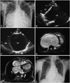
A 53-year-old woman presented with progressive dyspnea to our outpatient clinic. She reported first shortness of breath one year prior to the current presentation. Her medical history was unremarkable and vital signs were stable. Her electrocardiogram demonstrated atrial fibrillation with slow ventricular response. A massive cardiomegaly, as evidenced by a cardiothoracic ratio of 0.95, was noted (Fig. 1A). Transthoracic echocardiography revealed a mitral valve area of 0.74 cm2 with a mild degree of mitral regurgitation. The left atrium (LA) was so large that it was not possible to fit it to the screen in its entirety (Fig. 1B and C). For further evaluation, a cardiac magnetic resonance imaging (cMRI) was performed and showed a huge LA with an end-systolic volume of 1,125 mL, resulting in the right atrium (RA) and right ventricle (RV) being significantly compressed. Even the left ventricle was also compressed by the enlarged LA against the anterior chest wall (Fig. 1D). RA and RV volume were 18 mL and 70 mL, respectively. Mitral valve replacement with bileaflet prosthetic valve was performed concomitantly with LA volume reduction surgery.1)2) Immediate postoperative course was uneventful. Postoperative cMRI performed at the 8 week follow-up visit after discharge showed a marked reduction of LA (250 mL) with a normalization of RA, RV (170 mL) and left ventricle sizes (Fig. 1E). One month after corrective surgery, her functional capacity had significantly increased from NYHA Fc IV to Fc II and the size of the cardiac chambers had normalized (Fig. 1F).
Figures and Tables
 | Fig. 1Comparion of cardiac chambers before versus after corrective surgery. A: massive cardiomegaly on preoperative chest film. B: parasternal long axis view of transthoracic echocardiography. C: apical four chamber view of transthoracic echocardiography D: preoperative cMRI. E: postoperative cMRI. F: postoperative chest film. Ao: ascending aorta, LA: left atrium, LV: left ventricle, RA: right atrium, RV: right ventricle, cMRI: cardiac magnetic resonance imaging. |
References
1. Tamura Y, Nagasaka S, Abe T, Taniguchi S. Reasonable and effective volume reduction of a giant left atrium associated with mitral valve disease. Ann Thorac Cardiovasc Surg. 2008. 14:252–255.
2. Kim HJ, Shin DG, Hong GR, et al. Effect of left atrial decompression by percutaneous balloon mitral commissurotomy on the atrial electrophysiologic properties. Korean Circ J. 2007. 37:208–215.


 PDF
PDF ePub
ePub Citation
 Print
Print


 XML Download