

 PDF
PDF ePub
ePub Citation
 Print
Print


Introduction
The assessment of left ventricular (LV) systolic function is of vital importance because it is closely associated with future patient outcomes.1)2) Evaluation of LV systolic function is usually achieved by measuring the ejection fraction (EF) and fractional shortening (FS). Albeit useful, the EF and FS obtained by conventional approach are derived from endocardial movement. As such, they cannot reliably reflect the "actual" systolic function in the setting of increased LV wall thickness.3) In fact, LV systolic function measured by conventional EF or endocardial FS (eFS) in patients with LV hypertrophy is estimated to be almost always normal or supernormal in spite of the adverse prognosis of patients with LV hypertrophy.4) This paradoxical trend may be explained by the followings: first, the inner layer of the LV that includes the endocardium moves inward, further than the outer layer of the LV. Second, the inner layer of the "thickened" LV tends to show more inward movement during systole, compared to the inner layer of the LV with "normal" thickness.5) Therefore, it has been recommended that the LV midwall FS (LV-FSmw) be used in place of conventional eFS in patients with increased LV wall thickness for the assessment of LV systolic performance.6) From a prognostic point of view, LV-FSmw has proven its value as a predictor of cardiac death and cardiovascular morbidity in patients with arterial hypertension.6)7) Any general use of LV systolic functional parameters should be preceded by determining their normal reference ranges. As the number and proportion of older individuals progressively increase in the general population, knowledge of the normal reference ranges and the physiological changes associated with aging will become important. Although the normal reference ranges for LV-FSmw have previously been established by studies in the Western population,8)9) the reference values and physiological changes with age have yet been explored in the Asian populations.
The objective of this study was to determine the normal reference values and physiologic changes of LV-FSmw, based on 160 clinically normal Koreans and utilize the outcomes to establish reference standards for the assessment of LV-FSmw in patients with pressure or volume overload in future studies.
Subjects and Methods
Study population
Volunteers who visited the Health Care Center at our hospital for general routine check-up without laboratory abnormalities, including electrocardiographic and echocardiographic findings, were considered candidates for this study. Patients with a history of cardiovascular or systemic diseases were excluded. For inclusion, subjects less than 40 years of age had to undergo a modified Bruce protocol-based treadmill test and those beyond 40 years of age required a treadmill test and a computed tomographic coronary angiography (CTCA) in order to exclude subclinical coronary artery disease. Volunteers who had stenosis of >25% in any coronary artery on the CTCA and who did not reach at least 85% of age-predicted maximal heart rate during the treadmill test were systematically excluded. Subjects who were ≥50 years of age were screened by carotid Doppler examination and patients who had stenoses greater than 50% of the lumen diameter in any carotid arteries were excluded. The study protocol was approved by the institutional review board of our hospital and written informed consent was obtained from all participants before study enrollment.
Echocardiographic analysis
Echocardiographies were performed by one cardiologist using commercially available equipment (GE Vivid 7, GE medical systems, Milwaukee, WI, USA) with subjects in the left lateral decubitus position. LV cavity size, LV mass, LV relative wall thickness (RWT) and FSmw were determined using tissue harmonic imaging with M-mode measurements according to the guidelines suggested by the American Society of Echocardiography.10) Interventricular septal wall thickness (IVST), LV internal diameter (LVID) and posterior wall thickness (PWT) were measured at end-diastole and at end-systole. LV mass was calculated with the Devereux formula.11) To establish the relationship between LV mass and linear height (Ht), LV mass was adjusted for height by dividing the value by the Ht2.7.12) LV-RWT was calculated using (LV-IVSTd+LV-PWTd)/LVIDd. The LV-eFS was calculated using the conventional formula: e.g., LV-eFS (%)={(LVIDd-LVIDs)/LVIDd}×100, where s denotes end-systole and d, end-diastole. LV-EF was derived from modified Simpson's method.
Left ventricular midwall fractional shortening calculation
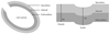
Calculation of LV-FSmw was performed with the assumption of a cylindrical shaped LV, including consideration of epicardial migration of midwall circumferential fibers during systole.13) The model that assumed LV geometry to be cylindrical shaped considered LV as the union of two concentric cylindrical shells, with equal thickness at end-diastole (Fig. 1). This model is based on the fact that LV wall volume is constant during the cardiac cycle. Using this model, the LV-FSmw can be calculated with the following formula: LV-FSmw={(LVIDd+LV-IVSTd/2+LV-PWTd/2)-(LVIDs+Hs/2)}/LVIDd+LV-IVSTd/2+LV-PWTd/2)
Stress-corrected left ventricular midwall fractional shortening calculation
In order to eliminate the effects of LV afterload on LV-FSmw, stress-corrected LV-FSmw was calculated by normalizing LV-FSmw to LV circumferential end-systolic wall stress (LV-cESS) in each subject.14) LV-cESS was calculated from the following formula: LV-cESS={[SBP×(LVIDs/2)2]×[1+(LVIDs/2+LV-PWTs)2/(LVIDs/2+LV-PWTs/2)2]}/{(LVIDs/2+LV-PWTs)2-(LVIDs/2)2}, where SBP is the brachial cuff systolic blood pressure (BP).
Statistical analysis
All measurements were analyzed by one observer using Statistical Package for the Social Sciences (SPSS) statistical software, version 14.0 (SPSS, Inc., Chicago, IL, USA). Data are presented as the mean±SD for continuous variables and number (percentages) for categorical variables. To minimize the impact of confounding factors due to the increased proportion of women in the older age group, partial correlation and analysis of covariance taking gender into account were performed. A p<0.05 was considered statistically significant.
Results
Clinical and conventional echocardiographic data
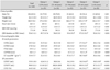
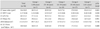
The study population was comprised of 160 subjects {104 males, 56 womens, age 44.9±2.7 years (range, 11-72 years)}. Clinical and conventional echocardiographic results across age deciles are summarized in Table 1.
Comparison of left ventricular-endocardial fractional shortening and left ventricular midwall fractional shortening without gender breakdown
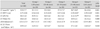
Table 2 shows the comparison between eFS and FSmw, with differentiation by age deciles in conjunction with data for the entire cohort. LV mass/Ht2.7 and LV-RWT showed significant increases with age (p for trend=0.002 and 0.001, respectively). LV-eFS and LV-EF also displayed a gradual increase with age (p for trend=0.001 and 0.002, respectively), whereas LV-FSmw and SC LV-FSmw remained constant, regardless of age (p for trend=0.88 and 0.29, respectively).
Comparison of left ventricular-endocardial fractional shortening, left ventricular midwall fractional shortening and stress-corrected left ventricular midwall fractional shortening according to gender
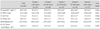
Echocardiographic findings of endocardial versus midwall mechanics by gender are shown in Table 3 and 4. Although the analyses were limited by small sample size, LV-RWT and LV-eFS showed significant increase, in parallel with age among men (p for trend=0.001 and 0.006, respectively) and women (p for trend=0.030 and 0.014, respectively). With advancing age, LV-FSmw tended to decrease, without statistical significance in men (p for trend=0.30). Despite a slightly higher LV-FSmw value in women compared to men (18.2±1.5% for male vs. 19.4±2.5% for female), this difference did not reach statistical significance (p=0.07). In the women, LV-FSmw showed no trend in the relationship with age (p for trend=0.99), as shown in Table 4. SC LV-FSmw value also showed no trend with age (p for trend= 0.29 for male and 0.50 for female).
Discussion
This is the first report on the normal reference values of LV-FSmw and the natural, physiological changes with age in a large number of healthy Korean individuals who underwent echocardiography using the harmonic imaging technique. In contrast to LV-eFS that showed a progressive increase in parallel with age, LV-FSmw displayed no significant changes in relation to aging in all subjects. This remained the case even when the analyses were performed to adjust for gender. Overall, the LV-FSmw values obtained in this study were comparable to that of previous studies.9)15) However, the trend for LV-FSmw change is different from that of previous studies, in which LV-FSmw decreased slightly with advancing age in normal subjects.9)15) This discrepancy can be explained by differences in the characteristics of the study population. Specifically, the subjects in the present study exhibited no increment in BP with age, whereas BP of the subjects enrolled in the study by Slotwinter et al.9) displayed stepwise BP increment in parallel with age, which can lead to a significant discrepancy between the two studies. In addition, silent myocardial diseases may account for some of the modest age-related decline in LV-FSmw observed in previous studies,15) which was not the case for our study due to systematic exclusion of subjects with subclinical coronary artery disease based on treadmill test and CTCA. We believe that this attempt is of crucial value, given that coronary artery disease is now the most common cause of subclinical myocardial dysfunction in Korea. In terms of conventional echocardiographic parameters, LVIDs, LVIDd, LV-IVSd, and LV-PWTd values in patients less than 50 years are comparable to that obtained in previous studies, albeit there exist discrepancies in subjects aged more than 50 years.16) However, the differences are very small and can be considered within a variability range that may be observed in routine echocardiographic examinations.
Echocardiography is a non-invasive and readily available diagnostic modality that performed in patients with known or suspected cardiovascular disease. Thanks to its versatility, the indications for echocardiography have progressively expanded. The assessment of LV systolic function is one of the most important clinical applications of echocardiography. Traditionally, echocardiographic evaluation of LV systolic function has been predicated using indexes acquired by measurements at the endocardial level. Many previous studies have shown that assessment of the LV systolic function based on endocardial movement affected the choice and timing of cardiac surgery and long-term survival.17)18) However, LV systolic function obtained on the basis of endocardial movement is of limited utility in patients with LV hypertrophy, given the reciprocal change of LV-EF in relation to LV wall thickness.19)20) In other words, the presence of ventricular hypertrophy can lead to over-estimation of LV-EF, the most frequently used index for estimating LV systolic performance, despite decline in myocardial function, as demonstrated by tagging magnetic resonance imaging.21) This apparent discrepancy could be explained, at least in part, by "cross-fiber shortening", where endocardial shortening tends to be greater than normal in patients with LV hypertrophy.22) Because subendocardial fibers are longitudinally arranged, circumferential and radial shortening observed at the endocardial level cannot be directly generated by shortening of subendocardial fibers per se.
To overcome the limitation of evaluating LV systolic function in patients with thickened LV walls, direct estimation of LV-FSmw was introduced and found to be physiologically more appropriate than LV-eFS in estimating LV systolic performance when LV hypertrophy was present.3)6) Furthermore, LV-FSmw was shown to be an independent risk factor to predict cardiovascular events in patients with hypertension.23-26) As the number and proportion of older individuals in the population progressively increases, the prevalence of hypertensive patients is expected to increase steadily and thus the utility of LV-FSmw could be augmented. For widespread use of LV-FSmw in the clinical arena, knowledge of its normal reference range and physiologic changes associated with aging become more and more pivotal.
This study, for the first time, demonstrated the normal reference ranges of LV-FSmw and its physiological changes with age in the Asian population. Cardiovascular disease states can only be characterized in the aging populations with reference to age-adjusted normal values. However, in terms of defining normal reference ranges, the sole effect of aging on natural remodeling of the heart is difficult to evaluate, because of subclinical diseases not infrequently found in clinical practice incidentally. Because all individuals in the present study were confirmed to be clinically normal based on laboratory tests, clinical and physical examination findings, CTCA and carotid Doppler studies, we believe that our study is free of inadvertent enrollment of patients with subclinical disease.
It is interesting to note that in the current study, LV-FSmw obtained in male subjects showed slightly higher values compared to the females, although the difference did not reach statistical significance (19.4±2.5% vs. 18.2±1.5%, p=0.068). In addition, while LV-eFS increased in a stepwise manner with age, LV-FSmw did not significantly vary by the age deciles, suggesting that age per se does not necessarily needed to be considered when interpreting LV-FSmw, which is useful in the clinical application of LV-FSmw when comparison is made between groups with different ages. This result is also supported by the fact that no changes were observed in stress-corrected LV-FSmw with age, which was performed to eliminate the influence of LV afterload on LV-FSmw.
There were several limitations to this study. First, our data was derived from a single center and they may not be directly applicable to general populations. Normal echocardiographic range of any specific parameter population should be derived from a random sample of the general population. In this respect, our population may not reflect the general population. However, we selected healthy subjects among the population who visited our health care center for general check-up at their discretion. Therefore, we believe that the present study population can be, to some extent, a representative sample of the Koreans.
In conclusion, the results of this study provide normative data for LV-FSmw and its physiological changes with age in a relatively large number of normal Asian subjects. LV-FSmw remained stable with age, which was in contrast to LV-eFS that showed progressive increase in parallel with age. The information obtained in the present study can be applicable to future studies, and provides reference standards with which LV systolic dysfunction can be defined more accurately.
 XML Download