

 PDF
PDF ePub
ePub Citation
 Print
Print


Introduction
The increasing incidence of complex percutaneous coronary intervention (PCI) is accompanied by risks of device fracture or dislodgement. Guide wire fractures during PCI are very rare, but in such cases, life-threatening complications such as embolization, thrombus formation and perforation may occur. Although, in most cases, percutaneous retrieval techniques of fractured guide wires are recommended, there have been several reports of fragments being left in place without complications.1-4)
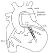
We present the case of a 78-year-old female patient who was diagnosed with non-ST segment elevation myocardial infarction (NSTEMI) and treated with PCI. The patient had remnant guide wire filaments in the left anterior descending artery (LAD) and aorta but did not experience any serious complications during a one-year follow up period.
Case
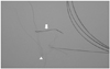
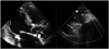
A 78-year-old woman presented with NSTEMI. Echocardiography showed decreased left ventricular systolic function (ejection fraction 45%). A diagnostic coronary angiogram showed diffuse significant stenosis from the proximal to middle LAD and first diagonal branch (D1) (Fig. 1). There was total thrombotic obstruction at the middle right coronary artery. We initially treated the patient with a single paclitaxel-eluting stent (2.75×32 mm Taxus®, Boston Scientific, Natick, MA, USA). Four days later, two coronary angioplasty 0.014 guide wires were inserted {one hi-torque Balance Middleweight (BMW) universal coronary guide wire (Abott Vascular, Santa Clara, CA, USA) into the LAD, and one high-torque Whisper coronary guide wire (Abott Vascular) into the D1}. An intravascular ultrasound after predilatation with a 2.5×20 mm Voyager® balloon (Abott Vascular) revealed a large plaque burden at both the LAD and the D1. We deployed two overlapped sirolimus-eluting stents at the proximal and middle LAD: a 2.75×33 mm Cypher® at the middle LAD and a 2.75×18 mm Cypher® at the proximal LAD (Cordis Corporation, Miami Lakes, FL, USA). When we exchanged the guide wires to perform kissing balloon angioplasty, a fracture occurred at the distal tip of the BMW guide wire (Fig. 1). Another guide wire was inserted to perform a beaded wire rotation, and distal balloon inflation retrieval was attempted, in order to remove the fractured guide wires. This attempt, however, was ineffective. Finally, we used a goose neck loop-snare (Microvena Corporation, St. Paul, MN, USA) to remove the fractured guide wires. Multiple forward and backward movements of the snare, combined with distal balloon inflation retrieval, successfully removed most of the fractured guide wires (Fig. 2) but we later observed retained filaments during echocardiography (Figs. 3 and 4). The patient declined surgical intervention for removal of these stray filaments and was discharged from our hospital, with triple anti-platelet medication and no complications. The patient did not experience any thrombotic or embolic events and did not suffer from any subjective symptoms over the one year of clinical follow up.
Discussion
Guide wire fractures during PCI are very rare, occurring in approximately 0.1-0.2% of cases.5) Guide wire remnants could lead to life threatening complications such as thrombosis, emboli, and perforation. Therefore, in the event of failed percutaneous retrieval and persistent signs of ischemia, patients should be urgently referred for surgical intervention. There are several methods recommended for the management of fractured guide wires, including emergent surgery, loop snare removal, two- or three-wire rotation, stenting over the retained wire, and conservative treatment.6-8) Surgical extraction is strongly recommended in cases of protrusion of the guide wire into the ascending aorta.9-11) However, guide wire segments retained within the coronary circulation may remain benign for a long time, particularly if they are entrapped within a distal part of the vessel and do not have accompanying total coronary occlusions.3) Vascular endothelial cell covering over the guide wire fragments may render them immobile and non-thrombogenic.
Hi-torque BMW guide wires consist of a distal core and a stainless proximal shaft, facilitating treatment of multiple lesions and tortuous vessels. However, guide wire fractures may occur if the distal core and stainless proximal shaft are separated by either the trapping of the distal tip or by vascular resistance. Because the fracture in the LAD developed after stent deployment at the main branch, we could not determine the mechanism of fracture in this particular case. However, trapping of the distal tip of the BMW wire, or stent deployment over a severely angulated guide wire are two possible explanations.
In conclusion, even though the most ideal management option for remnant guide wires is their removal, conservative treatment with the fragments left in situ may be successful in cases in which patients remain asymptomatic and hemodynamically stable. However, life-long administration of intensive anti-platelet medications and close observation are recommended for these patients.

 XML Download