

 PDF
PDF ePub
ePub Citation
 Print
Print


Introduction
Heart failure (HF) affects 4 to 5 million people in the United States, and more than 500,000 new cases are diagnosed annually.1) Although HF may strike at any age, it is more common in people over 65 years of age. HF not only affects systolic function, but also diastolic function either predominantly or in isolated manner.
Myocardial contraction and relaxation are active, energy-dependent processes.
Recently, there have been rapid advancements in the evaluation of myocardial function with non-invasive techniques,2) particularly magnetic resonance (MR) imaging has the potential to provide an insight into the pathophysiology of various heart diseases.3)4)
MR spectroscopy demonstrates the energy status of human cardiac tissue in vivo. The change in proton density on MR spectroscopy (1H-MRS) has been associated with disturbed myocardial function. 1H-MRS was therefore proposed as a non-invasive technique for the investigation of cardiac metabolism in humans, which thus far has been extensively used to examine myocardial metabolism in animals in vivo.5-7) Studies in human hearts have also been reported.8-12)
The aging process has been known to be associated with impaired energy metabolism of the heart. Exercise training in patients with congestive heart failure (CHF) provides physiological benefits and makes positive impact on the quality of life.13) However, it is unclear how exercise training affects the important aspects of myocardial metabolism in the aged heart. Therefore, in this study, we aim to utilize H-MRS to determine whether long-term exercise training will improve age-related myocardial metabolic derangement.1)
Materials and Methods
Animals
Old control (OC, 22 months of age) and young control (YC, 3 months of age) male Fischer 344 rats were obtained (Samtako, Osan, Korea). Three rats were housed per cage and fed with standard laboratory diet and water, and maintained on a 12:12-h alternate light-dark cycle. All rats were given one week to adjust to their new surroundings after arrival. No training was performed during this period. Body weights were recorded at baseline and subsequently weekly, as well as at the time of acquiring MR images.
Old rats were randomly assigned into sedentary control (OC), or exercise training {old exercise-trained (OT)}. Eventually 13 rats in YC, 15 rats in OC and 15 rats in OT were allocated. The rats were studied at the age of 6 months (young) and 25 months (old) after 12 weeks of exercise training or normal sedentary cage life. These investigations received the approval of the Seoul National University Hospital Institutional Animal Care and Use Committee.
Exercise training protocol
Before training commenced, all rats were familiarized with the treadmill by walking/running for 10 min/day for 10-15 days. The old exercise training groups commenced training at 4 m/min for 20-30 minutes, 5 days per week. The rate and time were progressively increased throughout the 12-week training program. The terminal rate was 15 m/min for 45-60 minutes/day, 5 days per week. These speed-grade combinations have been shown to be slightly less than 70-75% of the maximum oxygen consumption (VO2max).14)15) YC, OC rats walked or ran on the treadmill for 10 minutes per week to maintain familiarity with the treadmill for subsequent investigations of the maximum exercise capacity.
Maximal exercise capacity
At the end of the 12-week period, maximum exercise capacity was measured twice in each rat in a test consisted of walking at 10 m/min for 5 minutes followed by an increase in speed of 2 m per minute every 2 minutes, until the rat reached exhaustion.16)
In vivo 1H-MR spectroscopy
After 12 weeks of treadmill exercise training (6 months and 25 months of age, respectively), all spectroscopic experiments were carried out on an 4.7 T (200 MHz) MR system comprised of a vertical magnet (bore size 123 mm, Magnex Scientific, Oxon, UK), a Bruker Avance console (Bruker Medical, Ettlingen, Germany) and a shielded gradient system (548 mT/m, 160 µs rise time) (Magnex Scientific). Cardiac and respiratory signals were continuously monitored with electrocardiography and a respiratory gating device. Both signals were derived from two electrodes inserted subcutaneously in the front paws. Respiratory signals could also be obtained from a loop loosely fitted to the chest and the abdomen of the animals. Temperature was maintained using a blanket heated by warm air. Rats were secured within the holder using surgical tapes, without compressing their abdomen or chest. Surface coil with inner diameters of 37 mm were used for in vivo 1H-MRS. After positioning the rat in the magnet with the heart in the isocenter of the coil, seven contiguous, 1-mm thick axial slices were acquired using a segmented cardiac-triggered and respiration-gated FLASH-sequence. Selective shimming of a 3-4 mm3 volume located in the interventricular septum was performed manually using a PRESS sequence (TE=10-30 ms, TR=203 seconds), with a Shinnar-Le Roux optimized pulse for excitation and Mao-type refocusing pulses. The exact repetition time of the sequence was determined by the heart rate with physiologically derived steady state maintenance during respiration.17) Water-suppressed and unsuppressed cardiac spectra from a 2×2×3 mm voxel positioned in the septum were acquired in diastole using the same PRESS sequence. Water suppression was achieved by applying a CHESS module made up of three frequency-selective RF pulses (band width: 560 Hz) and doubling spoiler gradients. Water-suppressed spectra consisted of 256 averages (NA =256) were acquired on-resonance on creatine. In addition, to test the quality of motion compensation, an average was calculated in 128 consecutive spectra acquired in three of the rats without water suppression (NA=1). The experimental time was typically 1 minute per unsuppressed spectrum and 15 minutes per water-suppressed spectrum.
Data analysis of 1H-MR spectroscopy
Motion influence was quantitatively investigated by fitting each spectrum obtained from the repeated water signal measurements in Fischer rat. The two sets of water-suppressed spectra were added and fitted. The three spectra acquired without water suppression were also fitted, and the amplitudes of the metabolite peaks were normalized to the mean value of the three water peak amplitudes. Creatine signal was identified and measured at 3.0 ppm by the spectrometer's XWIN-NMR software. The mean creatine and water signal-line widths were 7 Hz and 19 Hz, respectively.9) Total myocardial creatine is calculated by multiplying tissue-water concentration with the ratio of the integrated signal area of creatine in the water-suppressed spectrum to that of water in the non-suppressed spectrum, with correction factors to account for spin-spin and spin-lattice relaxation effects as previously described.9)
Statistical analysis
Continuous variables are described as the means±standard error in each group. The Kruskall-Wallis test was used to compare metabolite measurements in the control and aging groups. Statistical Package for the Social Sciences (SPSS) version 13 (SPSS, Inc., Chicago, IL, USA) was used to perform statistical analyses. Probability values of <0.05 were considered statistically significant.
Results
Animal characteristics
No significant difference was observed in body weight at baseline among OC and OT groups (Table 1). At the end of the 12-week period, there was no difference in body weight between YC and OC. Left ventricle (LV) weight and the ratio of LV to body weight (an index of LV hypertrophy) were significantly increased in old rats compared with YCs, which reflects the aging process, whereas LV weight and the ratio of LV weight to body weight were not different between rats in OC and OT group after 12 weeks of exercise training.
In the event of death, an autopsy to ascertain the cause was not carried out. However, in most cases, the rats progressively lost weight, weakened and died. The tumors detected outside were developed in 33.3% of the aging rats before MR spectroscopy.
Exercise capacity
Exercise capacity was 14.7 minutes greater in OT than OC (20.1±1.9 minutes in OT, 5.4±2.3 minutes in OC; p<0.001). Twenty weeks of exercise training rendered old rats with a maximum exercise capacity that matched a group of untrained YC rats (17.9±1.5 minutes in YC, 20.1±1.9 minutes in OT; p>0.05).
MR spectroscopy
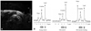
The creatine-to-water ratios in the interventericular septum of YC did not differ significantly from values obtained in OT (0.00131±0.00025 vs. 0.00127±0.00031; p=0.37). However, OC showed significant reductions in creatine-to-water ratio compared to OT (0.00096±0.00025 vs. 0.00127±0.00031; p<0.0). Therefore, OC is characterized by substantially reduced myocardial creatine levels, whereas OT maintained equivalent levels found in YC (Fig. 1).
Mean total creatine concentrations were similar in the myocardium of YC and OT (13.3±3.6 vs. 11.5±4.1 mmol/kg wet weight; p=0.29). In contrast, the mean total creatine concentration of OC showed a significant decline compared to OT (6.8±3.2 vs. 11.5±4.1 mmol/kg wet weight; p=0.03).
Discussion
In this study, we demonstrated that long-term exercise training in aged rats prevented age-related deterioration in myocardial metabolism. Long-term exercise training rendered old rats a maximum exercise capacity comparable to that of untrained YC rats. The creatine-to-water ratio and mean total creatine concentrations of myocardium in old trained rats did not differ from that in YC. In contrast, OC showed significant reductions in creatine-to-water ratio, and mean total creatine concentration, compared to old trained and YC rats.
Osbakken et al.18) suggested that changes in myocardial creatine kinase kinetics may contribute to myocardial dysfunction using 31P-MR spectroscopy. Spindler et al.19) also insist that the energy-requiring processes may contribute to the observed myocardial dysfunction, which is probably related to a calcium overload. Therefore, metabolic changes in MR spectroscopy have been associated with disturbed myocardial function.20)
Lipid metabolism is of special interest in the evaluation of myocardial status.21-23) In addition, invasive animal and human studies have indicated that the total creatine content is reduced in the failing heart.24)25) Creatine is critical for myocardial energy metabolism and reserve, and evidence suggests that reduced energy reserve underlies impaired contractile function in the failing heart. The direct, non-invasive detection and measurement of creatine with 1H-MR spectroscopy represents an exciting opportunity to probe myocardial metabolism in healthy and diseased subjects.26) Therefore, 1H-MR spectroscopy may be useful in the study of normal cardiac physiology, and in the diagnosis and monitoring of HF treatment.10)
In this study, we found that 12 weeks of exercise training in aged rats can improve creatine metabolism. The underlying molecular mechanism for this improvement could either be reversal of the age-associated changes that resulted in impaired myocardial function, or training in general conferred some beneficial effects on myocardial function. Our finding that exercise improved cardiac creatine metabolism in the aged rat probably represent the in vivo manifestation of improved energy-dependent calcium release and reuptake of the whole heart that others have reported in isolated muscle strips from trained, aged rat hearts.27)28) Therefore, from this study using 1H-MRS, we suggest that creatine metabolism may play a substantial causative role underlying the progression or improvement of HF.
However, there are limitations in our study. First, we did not perform 31P-MR spectroscopy. Brain and skeletal muscle tissue in animal model have previously been characterized by 1H- and 31P-MR spectroscopy in vivo. However, Schneider et al.7) verified that a lack of creatine on 1H-MR spectroscopy must also lead to phosphocreatine depletion on 31P-MR spectroscopy. However, this technique has been hampered by low intrinsic sensitivity and metabolite concentrations, which have restricted studies to large myocardial voxels (20 mL in clinical studies) near the anterior chest wall,28) and 31P-MRS does not provide access to unphosphorylated creatine. The high sensitivity of 1H-MRS means that the higher concentration of creatine, and the higher content of 1H in the creatine N-methyl resonance at 3.0 ppm, approximately confers a 20-fold net theoretical improvement in sensitivity compared with 31P-MRS of phosphorylated creatine.
Second, measurements were made under light sedation and therefore at heart rates that are likely to be physiologically relevant.

 XML Download