

 PDF
PDF ePub
ePub Citation
 Print
Print


Introduction
Thyrotoxicosis can be manifested by many cardiovascular symptoms including palpitation, exercise intolerance, dysrhythmia, systolic hypertension, cardiomegaly, peripheral edema, and angina.1) Over 90% of patients experience tachycardia, and 10% experience atrial fibrillation or atrial flutter. In addition, chronic or severe thyrotoxicosis can aggravate sinus tachycardia or atrial fibrillation and cause heart failure accompanied by left ventricular systolic dysfunction.1-3) Heart failures by this mechanism have been explained as tachycardia-induced cardiomyopathy in previous studies.4)
Recently, it has been reported that thyrotoxicosis is presented first as right ventricular failure accompanying pulmonary artery hypertension and tricuspid regurgitation.5) However, most studies on right ventricular dysfunction (RVD) related to thyrotoxicosis mention only isolated right ventricular failure,6-11) and there are almost no studies on about wheather both left ventricular dysfunction (LVD) and right ventricular failure occur simultaneously, or there is correlation between the two. Therefore, to the purpose of this study was to identify factors that affect right ventricular systolic dysfunction occurring in thyrotoxicosis, and particularly examine whether RVD is related to LVD by tachycardia-induced cardiomyopathy occurring in thyrotoxicosis patients.
Subjects and Methods
Subjects
This research is a retrospective study on 12 patients diagnosed with thyrotoxicosis and LVD {left ventricle ejection fraction (LVEF) <40%}. Among the patients (male:female=3:9, mean age=51±11) that were admitted to Kangdong Sacred Heart Hospital of Hallym University College of Medicine for heart failure between September 2005 to October 2007, 9 were diagnosed with thyrotoxicosis for the first time and 3 were diagnosed in previous years, but not treated. In addition, coronary artery disease, hypertension, valvular heart disease, drug use, and infection were ruled out as causes of LVD.
Methods
The 12 patients diagnosed with thyrotoxicosis and LVD were divided into a group (group 1) with RVD and a group (group 2) without RVD by using tricuspid annular plane excursion (TAPSE). The TAPSE estimated RV systolic function by measuring the level of systolic excursion of the lateral tricuspid valve annulus towards the apex in the four chamber view. If the measured TAPSE was 15 mm or shorter, a patient was was defined as having RVD (group 1), and if it was 15 mm or longer, the patient was defined as non-RVD (group 2).
The sex, age, and clinical conditions, including the presence of underlying diseases and heart rates, echocardiographic findings, blood tests including troponin, B-type natriuretic peptide (BNP), and thyroid function tests at the time of admission were compared between 2 groups. In addition, when the two groups achieved an euthyroid state after antityhroid therapy, the clinical condition, echocardiographic parameters and blood treats were compared between two groups.
Left atrial dimension, diastolic left ventricular dimension, LVEF, diastolic right ventricular dimension, maximum pressure difference of tricuspid regurgitation, and postcaval dimension were measured with the use of transthoracic echocardiography. As for left atrial dimension, maximal anteroposterior length was measured by M-mode echocardiography at the aortic level of the parasternal short axis view. Diastolic left ventricular dimensions were measured immediately before the QRS of an electrocardiogram with M-mode echocardiography at the parasternal short axis view. The right ventricular anteroposterior dimension was measured at the end of systole when right ventricular volume reached its maximum in the parasternal long axis view. While the inferior vena cava (IVC) dimension can be measured at a subcostal window, the widest length at exhalation was selected. Tricuspid regurgitation (TR) was classified into mild, moderate, or severe by measuring the degree of regurgitation. Mild TR is defined as when regurgitation past the tricuspid valve reaches 1/3 entire distance of right atrium but not 1/2; moderate TR as when the regurgitation reaches 1/2 the distance but not 2/3; and severe TR as when regurgitation reaches beyond 2/3 the distance, nearly to the rear wall of the right atrium.
Statistical analysis
For statistical analysis, Statistical Package for the Social Sciences (SPSS) 12.0 (SPSS Inc., Chicago, IL, USA) software for Windows was used. All data are presented in mean±standard deviation, and for discontinuous variables between the two groups, the chi square test and the Mann-Whitney test were used. In order to determine changes in clinical status, blood tests and echocardiographic indicators at the time of admission and after treatment of thyrotoxicosis, the Wilcoxon signed rank test was performed. Values were considered significant if the p was less than 0.05.
Results
Clinical characteristics (Table 1)
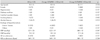
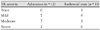
The mean age of all patients was 51±11, and there were more female (n=9, 75%) patients than male. Three patients had diabetes, hypertension or cerebrovascular disease; 3 patients smoked; and 2 patients had a history of alcohol use. Eleven patients showed atrial fibrillation with tachycardia, and the remaining patient showed sinus tachycardia with a heart rate of 150/min. The cause of thyrotoxicosis in 11 patients was Graves' disease, and 1 patient had suspected thyroid follicular adenomatosis. Six patients showed RVD (50%), and clinical characteristics between the two groups were not significantly different. Two patients underwent coronary angiography to rule out ischemic heart disease while hospitalized, and both belonged to RVD group. Neither had any lesions in the coronary artery, but one patient showed coronary vasospasm and the another showed myocardial bridging findings.
Blood test
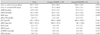
There was no significant difference found between the two groups in any blood tests obtained at admission, including free thyroxine (free T4), triiothyronine (T3), BNP, troponin, creatine kinase-MB (CK-MB) and C-reactive protein (CRP). Group 1 showed higher low density lipoprotein-cholesterol level compared with group 2, but the difference was not statistically significant (p=0.055). Serum creatinine values were significantly higher in group 1 (p<0.05), but mean value was within normal range (Table 2).
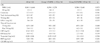
There was no significant difference between the two groups in tests for anti-thyroglobulin autoantibody, antithyroid peroxidase antibody (TPOAb) and thyroid stimulating hormone receptor antibody (TSHRAb) (Table 3).
Echocardiography (Table 4)
There were no statistically significant differences between the two groups in the results of echocardiography on the left atrial dimension, diastolic left ventricular dimension, LVEF, diastolic right ventricular dimension, the maximum pressure gradient of TR, or the IVC dimension. In the RVD group, the diastolic right ventricular dimension tended to be greater than normal group, but the difference was not statistically significant (p=0.078).
Results after antithyroid drug treatment
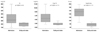
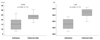
All patients took propylthiouracil (PTU) as antithyroid drug agent, and the free T4 and T3 values of all patients reached normal levels in an average of 85.8±70.5 days, this was a statistically significant change (Fig. 1). The 11 patients that showed atrial fibrillation with tachycardia showed a significant decrease of heart rate from 149.3/min to 75.1/min in average, and two patients were converted to normal sinus rhythm (Fig. 2). LVEF and TAPSE were improved over 40% and 15 mm respectively in an average of 61.1±66.5 days, also a statistically significantly increase (Fig. 2). TR was significantly improved as thyroid function returned to normal (Table 6). However no significant differences were found in blood pressure, BNP, diastolic left ventricular dimension, right ventricular dimension, the maximal pressure gradient of TR, or the IVC after treatment.
When the two groups were compared after restoration to normal thyroid function, clinical parameters including blood pressure and heart rate did not show a significant difference, nor did echocardiographic parameters, the thyroid function test (free T4, T3), BNP, left atrial dimension, LVEF, diastolic right ventricular dimension, the maximal pressure gradient of TR, or the IVC dimension (Table 5).
Discussion
Thyroid hormone (particularly T3) increases heart rate and left ventricular contractility, decreases systemic vascular resistance, increases sodium re-absorption and erythropoietin secretion, and raises stroke volume up to 300%.1) However, during thyrotoxicosis, atrial fibrillation or sinus tachycardia can occur, which may be caused by a calcium circulation distrubance to heart cells and structural changes in the heart.1-3) In case that atrial fibrillation or sinus tachycardia lasts for an extended period due to uncontrolled thyrotoxicosis, 6% of the patients experience heart failure, and among these cases half are accompanied by LVD; this is termed tachycardia induced cardiomyopathy.3)4)
Recently, a few studies have reported that thyrotoxicosis is manifested first by right heart failure with pulmonary artery hypertension and tricuspid regurgitation, but only isolated right ventricular failure was mentioned,7-12) and no study has been reported in which both RVD and LVD occur simultaneously. The purpose of this study is to determine the causative factors that trigger RVD in thyrotoxicosis and to examine the relationship between RVD and LVD in tachycardia induced cardiomyopathy.
Among the patients that experienced LVD by thyrotoxicosis, half (50%) had RVD. All 12 patients showed improvement in clinical features, blood tests, and echocardiography after thyrotoxicosis treatment. On admission, the RVD group (group 1) and the non-RVD group (group 2) did not show any significant differences in blood tests or clinical features including heart rate and BNP. Echocardiographic parameters also did not show any significant differences between the two groups.
Even in the euthyroid state, there was no significant difference between the two groups in the results of all tests, including changes in heart rate and the change of LVEF. That suggests that RVD occuring in thyrotoxicosis patients experiencing LVD has a different mechanism, compared with tachycardia induced cardiomyopathy known as mechanism of right heart failure in thyrotoxicosis.
If RVD in thyrotoxicosis is not caused by a secondary change in tachycardia-induced cardiomyopathy or secon-dary pulmonary artery hypertension, the right ventricle itself may be considered as the cause of RVD. Research indicating that the direct application of thyroid hormone to myocardial cells causes toxic effects has been explained by myocardial stunning.13)14) In addition, Kiss et al.15) found that the function of calcium ions within myocardial cells is deteriorated in thyrotoxicosis patients, and demonstrated that thyroid hormone affects transcription within myocardial cells and inhibits the synthesis of required proteins, triggering heart failure.16) In practice, thyroid hormone is known to affect many ion channels in the myocardial cell membrane, as well as gene expression within the nucleus of the myocardial cell.2) However, as thyroid hormone affects all myocardial cells and is not specific to the right ventricle, this data is not sufficient to explain the cause of RVD dysfunction. We hypothesize that when exposed to thyroid hormone, sensitivity of thyroid hormone receptor (TR) isomers TRα1, TRα2, and TRβ1 within myocardial cell changes; this mayalter the effects of thyroid hormone and explain the mechanism of RVD.1)2)
The limitation of this research is that we only studied 12 patients, so it is hard to draw statistically significant conclusions. However, only few case reports have been presented on thyrotoxicosis and right heart failure, and even in the largest study, performed by Cohen and Schattner7) only 8 patients were examined. In addition, since this is a retrospective study, cardiac catheterization for the accurate measurement of pulmonary artery pressure was not performed.
In conclusion, we demonstrate that RVD occurring in thyrotoxicosis is not related to secondary hemodynamic change by left ventricular systolic dysfunction. We hypothesize there may be a direct toxic effect of thyroid hormone on right ventricle, but further study is needed to validate this idea. However, we believe it is reasonable given that thyroid hormone affects gene expression in the nuclear of myocardial cells and works on various ion channels existing in the myocardial cell membrane. A sensitivity difference to thyroid hormone within cells could affect right heart failure. I think it is needed further study about molecular biology.


 XML Download