

 PDF
PDF ePub
ePub Citation
 Print
Print


Introduction
Mitral annular calcification (MAC) is a chronic, fibrotic degenerative change of the annulus supporting the mitral value.1)2) Histopathologically, MAC has been reported to occur when foam cells, which are characteristic of early-stage atherosclerotic lesions, are deposited on the epithelium lining of cardiac vessels, especially on the posterior mitral leaflet.3) MAC has been demonstrated to correlate with age,4) endocarditis,5) cardiovascular disease,6-10) and congestive heart failure.4)11)12) Furthermore, MAC has also been reported as a predictor of cardiovascular mortality.6)13)14)
With the aging of the current population, the number of elderly people aged 65 years or older has rapidly increased.15) The number of cases in which elderly people aged 90 years or older are hospitalized has also been increasing. However, few studies have examined the occurrence of MAC in these elderly patients.
Therefore, the objective of this study was to examine the clinical significance of MAC associated with the nonagenarian when compared with a group of young control patients.
Subjects and Methods
Study population
The current study was conducted in 52 elderly patients aged 90 years or older who were hospitalized to undergo cardiac echocardiography at the Division of Cardiology during a 3-year period from June 2005 to July 2008. The control group comprised 51 consecutive patients, from 20 to 50 years old, who underwent echocardiography. In the nonagenarian group, five patients had medical records regarding their echocardiography which were obscure and two had missing medical records. Total of 45 patients were finally enrolled in the nonagenarian group.
Measurement of mitral annular calcification
Medical records were evaluated for 96 patients. Clinical data of past medical history including hypertension, diabetes mellitus, hyperlipidemia, myocardial infarction, and family history of cardiovascular diseases were collected. Echocardiography was performed based on the judgment of physicians and using a GE Vivid 7 Ultrasound (General Electric Healthcare, Vingmed, Horten Norway) with a 2.5 MHz probe. The diameter of the left atrium, the size of the ascending aorta, left ventricular (LV) end-diastolic volume, LV end-systolic volume, the thickness of the interventricular septum, the thickness of the posterior ventricular wall, E wave, and E' wave were measured. The LV volume was measured by two-dimensional (2D) echocardiography and the left ventricular ejection fraction (LVEF) was measured using modified Simpson method.16) LV mass and LV mass index were calculated. LV mass were calculated using Devereux's formula:17)
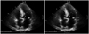
The LV mass index was defined as the LV mass, obtained using the formula above, divided by the body surface area. MAC was defined as an echo-dense structure observed in the margin of the atrioventricular groove and the posterior leaflet on the apical four chamber view and the parasternal long-axis view on a 2D echocardiography or an echo-dense structure observed posterior to the posterior leaflet on the parasternal short-axis view. This was confirmed by performing echocardiography for at least three cardiac cycles, and MAC width was then determined by selecting the thickest value of the measurements obtained from the apical four chamber view of 2D echocardiography (Fig. 1).8)14)
Statistical analysis
All the measurements were expressed as mean±SD. An inter-group comparison was made using Student's t-test. For MAC, the correlation of each variable was evaluated using a simple linear regression analysis. Statistical analysis was performed using Statistical Package for the Social Sciences (SPSS Inc. Chicago, IL, USA) version. A p<0.05 was considered statistically significant.
Results
Clinical characteristics
Of the nonagenarian patients, 12/45 were men (27%) and 73% were women (33/45) and the mean age was 92±2 years. Of the young control group patients, 26/51 were men (51%) and 49% were women (25/51) and mean age was 36±9 years.
The incidence of hypertension was higher in the nonagenarian patients {37/45 (82%)} than young control group {8/51 (15%), p<0.0001}. However, the incidence of smoking was higher in patients in the young control group {12/51 (54%)} than in the nonagenarian group {5/45 (24%), p=0.025}. There were no significant differences in diabetes mellitus (p=0.123), hyperlipidemia (p=0.938), past history of myocardial infarction (p=0.123), or family history of cardiovascular disease (p=0.097) between the two groups. Of the cardiovascular diseases, the incidence of heart failure was higher in nonagenarian {18/45 (40%)} than in young control group {6/51 (12%), p=0.0012} (Table 1).
Comparisons of the echocardiographic parameters
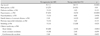
In comparing the echocardiographic results of the nonagenarian group with those of the young control group, significant increases were seen in the thickness of the interventricular septum (p<0.0001) and the thickness of the posterior wall of the LV in the nonagenarian group (p<0.0001). LV mass was also increased in elderly patients (165.79±49.34 vs. 145.67±40.02 gm, p=0.034), and the LV mass index was larger in nonagenarian patients when compared with the young patients (p<0.0001). The LV end-systolic diameter was similar between the two groups (p=0.725); however, the LV enddiastolic diameter was smaller in nonagenarian patients (p=0.007). The E/E' ratio was higher in nonagenarian patients (17.25±3.66 vs. 8.77±3.33, p<0.0001). Finally, the LVEF was significantly higher in the young control group (p=0.001) (Table 2).
Correlation between the clinical characteristics, echocardiograph parameters, and mitral annular calcification
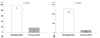
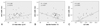
The incidence of MAC was 97% (41/43) in nonagenarian patients and 17% (9/51) in the young control group patients (p<0.0001). The width of MAC was 0.52±0.17 mm in nonagenarian patients and 0.05±0.13 mm in the young control group (p<0.0001) (Fig. 2). Simple linear regression analysis showed that the width of MAC correlated with age (r=0.836, p<0.0001), but not with the diameter of the left atrium (r=0.201, p=0.051), the LV end-systolic diameter (r=0.009, p=0.93), or the LV end-diastolic diameter (r=0.194, p=0.059). There were weak correlations between MAC and the thickness of the interventricular septum (r=0.363, p<0.0001), the thickness of the posterior wall of the LV (r=0.298, p=0.003), the size of the ascending aorta (r=0.382, p<0.0001), and LV mass (r=0.280, p=0.014) (Fig. 3A). The LVEF was negatively correlated with MAC (r=-0.340, p=0.001) (Fig. 3B), and the E/E' ratio showed a strong positive correlation with MAC (r=0.683, p<0.0001) (Fig. 3C). However, it was found that when adjusting for age, the LVEF (r=0.0043, p=0.6802), LV mass (r=0.0381, p=0.2335), and E/E' ratio (r=0.1656, p=0.1676) were not correlated with MAC. In addition, following adjustment for gender and underlying diseases, excluding cases in which a statistical significance could not be found due to a small number of enrolled patients, the correlation of the width of MAC with age was shown to be higher in nonagenarian patients (Table 3).
Discussion
The proportion of elderly people in Korea aged 65 years and older was 6.6% of the total population in 1998 and approximately 10.3% of the population in 2008. This indicates that the Korean population is aging rapidly.15) Cardiovascular disease is known to be the third most prevalent cause of death, after cancer and cerebrovascular disease, in elderly Koreans and shows a similar profile of incidence. However, few studies have reported the risk of cardiovascular disease in elderly Koreans.
According to Sadig et al.18) left ventricular hypertrophy (LVH) was markedly observed in elderly patients aged 100 years or older. In these patients, the LV diameter and LV end-systolic volume were significantly decreased, but the LVEF was higher than in young patients, which is thought to be a paradoxic response followed by decrease of LV volume and progression of LVH. Wong et al.19) reported that the thickness of the interventricular septum, the thickness of the posterior wall of LV, LV mass, and LV mass index were increased in women aged 50 years or older. In the study presented here, the size of the aorta, the thickness of the posterior wall of the LV, the thickness of the interventricular septum, LV mass, LV mass index, and the ratio of E/E' were significantly increased in elderly patients aged 90 years or older, which is consistent with the previous studies. Additionally, the LVEF was maintained within the normal range in these elderly patients, whereas the LVEF exceeded normal range (55-75%) in elderly patients aged 100 years or older.18)
It has been reported that mitral stenosis is associated with the calcification of the mitral valve20) and coronary artery,21-24) a giant aneurysm of the Sinus of Valsalva with calcification,24) calcification of left atrium,25) and aortic stiffness.26) However, reports of MAC are limited. Boon et al.27) conducted a comparative study in 657 patients with MAC; dividing patient groups as under 45 years of age, between 45 and 55 years of age, between 56 and 65 years of age, and over 66 years of age; and found that the increase in MAC was correlated with age. Rao et al.28) reported that the LVEF decreased depending on the severity of MAC in patients with chronic renal failure, and this led to the impairment of the contractile function of LV. In addition, in cases in which the ratio of E/E' was smaller than 10, pulmonary artery wedge pressure (PAWP) was lower than 15 mmHg, and when the ratio of E/E' was greater than 20, PAWP exceeded 20 mmHg. These results suggest that PAWP is associated with the increase in LV filling pressure.29)30)
In this study, the frequency and width of MAC were shown to be significantly higher in nonagenarian patients. After adjusting for gender and underlying diseases, the width of MAC was significantly higher in these elderly patients, suggesting that MAC is an indicator of the degenerative changes of the heart associated with age. Additionally, MAC appears to be an indicator of increased LV filling pressure, as is the ratio of E/E' in elderly patients.
This study was a small retrospective study. We could not evaluate the correlation of cardiovascular risk factors such as hypertension, diabetes mellitus, hyperlipidemia, and smoking with MAC because of the limited use of 2D echocardiography in nonagenarian patients. Also, the differences between male and female patients were not analyzed. The quantitative measurement of MAC was limited using 2D echocardiography. More importantly, MAC itself diminished the motion of the mitral annulus, so the assessment of the E/E' ratio did not reveal the actual LV filling pressure.
MAC is a degenerative change that occurs in elderly patients, and is associated with the LV filling pressure. Further studies are warranted to examine the structural changes of the heart and the risk of cardiovascular mortality associated with MAC.


 XML Download