

 PDF
PDF ePub
ePub Citation
 Print
Print


Abstract
Background and Objectives
P-wave dispersion (PWD) is a well-known electrophysiologic parameter of atria which are prone to fibrillation. Although paroxysmal atrial fibrillation (PAF) following an acute myocardial infarction (AMI) is not uncommon, the relationship between PWD and PAF following AMI has not been determined.
Subjects and Methods
We reviewed the electrocardiograms, recorded on admission and every day during hospitalization, of 144 patients with primary anterior AMIs and measured the P-wave duration. The left atrial diameter and left ventricular ejection fraction (LVEF) were evaluated by echocardiography.
Results
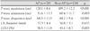
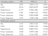
PAF occurred in 20 patients. The maximum P-wave duration and PWD were found to be significantly higher in patients with PAF than those without PAF (120.1±8.6 vs. 109.2±12.2 ms, p<0.001; and 68.5±11.9 vs. 48.7±9.6 ms, p<0.001, respectively). The minimum P-wave duration was significantly lower in patients with PAF than in patients without PAF (51.6±13.3 vs. 60.4±11.7 ms, respectively, p=0.003). There was no significant difference in the left atrial diameter between patients with PAF and patients without PAF (37.3±4.4 vs. 36.8±5.1 mm, respectively p=0.652); however, the LVEF was significantly different in the patients who developed PAF compared to those who did not develop PAF (38.5±11.4 vs. 45.1±8.7%, respectively, p=0.003).
Figures and Tables
Fig. 1
Correlation between P-wave dispersion and left ventricular ejection fraction. A: group without atrial fibrillation. B: group with atrial fibrillation. r: correlation coefficient, CI: confidence interval.
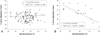
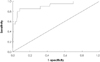
Fig. 2
Receiver operating characteristics (ROC) curve of P wave dispersion {area under the curve (AUC), 0.908}.
References
1. Kim DH, Kim GC, Kim SH, et al. The relationship between the left atrial volume and the maximum P-wave and P-wave dispersion in patients with congestive heart failure. Yonsei Med J. 2007. 48:810–817.
2. Kose S, Aytemir K, Can I, et al. Seasonal variation of P-wave dispersion in healthy subjects. J Electrocardiol. 2002. 35:307–311.
3. Olzer N, Aytemir K, Altalar E, et al. P wave dispersion in hypertensive patients with paroxysmal atrial fibrillation. Pacing Clin Electrophysiol. 2000. 23:1859–1862.
4. Dilaveris PE, Andrikoupoulos GK, Metaxas G, et al. Effects of ischemia on P wave dispersion and maximum P wave duration during spontaneous angina episodes. Pacing Clin Electrophysiol. 1999. 22:1640–1647.
5. Lee CK, Kim DH, Kim GC, et al. The role of P-wave from surface electrocardiography for the prediction of atrial fibrillation after coronary bypass graft surgery. Korean Circ J. 2005. 35:677–682.
6. Dilaveris PE, Gialafos EJ, Sideris SK, et al. Simple electrocardiographic markers for prediction of paroxysmal idiopathic atrial fibrillation. Am Heart J. 1998. 135:733–738.
7. Kim W, Shin DG, Hong GR, Park JS, Kim YJ, Shim BS. Signal averaged P wave dispersion: a new marker for predicting the risk of paroxysmal atrial fibrillation. Korean Circ J. 2002. 32:339–348.
8. Hod H, Lew AS, Keltai M, et al. Early atrial fibrillation during evolving myocardial infarction: a consequence of impaired left atrial infarction. Circulation. 1987. 75:146–150.
9. Eldar M, Canetti M, Rotstein Z, et al. Significance of paroxysmal atrial fibrillation complicating acute myocardial infarction in the thrombolytic era. Circulation. 1998. 97:965–970.
10. Pedersen OD, Bagger H, Kober L, Torp-Pedersen C. The occurrence and prognosis significance of atrial fibrillation/flutter following acute myocardial infarction. Eur Heart J. 1999. 20:748–754.
11. Goldberg RJ, Seeley D, Becker RC, et al. Impact of atrial fibrillation on the in-hospital and long-term survival of patients with acute myocardial infarction: a community-wide perspective. Am Heart J. 1990. 119:996–1001.
12. Behar S, Zahavi Z, Goldbourt U, Reicher-Reiss H. Long-term prognosis of patients with paroxysmal atrial fibrillation complicating acute myocardial infarction. Eur Heart J. 1992. 13:45–50.
13. Choi WG, Kim DH, Kim KC, et al. Change of the P wave duration and P wave dispersion according to treatment strategy in patients with a acute myocardial infarction. Korean J Med. 2007. 73:489–495.
14. National Cholesterol Education Program (NCEP) Expert Panel on Detection, Evaluation, and Treatment of High Blood Cholesterol in Adults (Adult Treatment Panel III). Third Report of the National Cholesterol Education Program (NCEP) Expert Panel on Detection, Evaluation, and Treatment of High Blood Cholesterol in Adults (Adult Treatment Panel III) final report. Circulation. 2002. 106:3143–3421.
15. Mahaffey KW, Granger CB, Sloan MA, et al. Risk factors for in-hospital nonhemorrhagic stroke in patients with acute myocardial infarction treated with thrombolysis. Circulation. 1998. 97:757–764.
16. Andrikopoulos G, Dilaveris PE, Richter DJ, Gialafos EJ, Synetos AG, Gialafos JE. Increased variance of P wave duration on the electrocardiogram distinguishes patients with idiopathic atrial fibrillation. Pacing Clin Electrophysiol. 2000. 23:1127–1132.
17. Aytemir K, Ozer N, Atalar E, et al. P wave dispersion on 12-lead electrocardiography in patients with paroxysmal atrial fibrillation. Pacing Clin Electrophysiol. 2000. 23:1109–1112.
18. Myrianthefs MM, Shandling AH, Startt-Selvester RH, et al. Analysis of the signal-averaged P wave duration in patients with percutaneous coronary angioplasty-induced myocardial ischemia. Am J Cardiol. 1992. 70:728–732.
19. Rios JC, Schatz J, Meshel JC. P wave analysis in coronary artery disease: an electrocardiographic-angiographic and hemodynamic correlation. Chest. 1974. 66:146–150.
20. Celik T, Iyisoy A, Kursaklioglu H, et al. The comparative effects of telmisartan and ramipril on P-wave dispersion in hypertensive patients: a randomized clinical study. Clin Cardiol. 2005. 28:298–302.
21. Tuncer M, Gunes Y, Guntekin U, Gumrukcuoglu HA, Eryonucu B. Short-term effect of cilazapril and atenolol on P-wave dispersion in patients with hypertension. Adv Ther. 2008. 25:99–105.
22. Crenshaw BS, Ward SR, Granger CB, Stebbins AL, Topol EJ, Califf RM. Atrial fibrillation in the setting of acute myocardial infarction: the GUSTO-I experience. J Am Coll Cardiol. 1997. 30:406–413.
23. Kim HJ, On YK, Sung JD, et al. Risk factors for predicting newonset atrial fibrillation in persons who received health screening tests. Korean Circ J. 2007. 37:609–615.
 XML Download