

 PDF
PDF ePub
ePub Citation
 Print
Print


Introduction
The pathophysiology of hypertrophic cardiomyopathy (HCM) is complex, with structural, hemodynamic, and arrhythmic processes contributing to the symptoms and natural history in this disorder. However, the most important pathophysiologic features of HCM is diastolic dysfunction and a decrease in left ventricular compliance. Thus, HCM is regarded as the most representative disease of left ventricular diastolic dysfunction.1)2) The histopathologic findings of HCM are myocardial hypertrophy, and interstitial fibrosis and disarray. Previous studies based on autopsies have shown that the degree of myocardial fibrosis is severe and varies as compared with myocardium obtained from normotensive or hypertensive patients.3) In recent years, several studies have been conducted to quantify the myocardial fibrosis and have reported that serum biochemical markers, which are synthesized by the production and degradation of matrix collagen, are elevated in subjects with hypertensive myocardial hypertrophy and HCM relating to the myocardial fibrosis.4-6) It has also been suggested that serum biochemical markers are associated with diastolic dysfunction.7-10)
The most common symptom of HCM is shortness of breath and it is usually aggravated by exercise.1) Diastolic dysfunction seems to be the most important determinant of exercise capacity in patients with HCM. In HCM, the limited capability of increasing left ventricular end-diastolic volume, especially during exercise at high heart rates, implies an inadequate increase in stroke volume.11) Such a lack of increase in stroke volume is accompanied by a leftward shift of the left ventricular diastolic pressure-volume relation during exercise.11)12) Specifically, based on a few studies which were conducted using exercise Doppler echocardiography, it has been demonstrated that left ventricular diastolic reserve, systolic reserve, and exercise tolerance are decreased in patients with HCM.13)14) However, there are many differences in exercise capacity and symptoms of heart failure, although there is a similar extent of myocardial hypertrophy or the left ventricular dimension in patients with HCM. Thus, we can hypothesize that the degree of myocardial fibrosis might be associated with the left ventricular reserve and exercise tolerance. To prove our hypothesis, we sought to assess diastolic function not only at rest, but also with exercise by using exercise Doppler echocardiography in patients with HCM. To quantify the amount of myocardial fibrosis, serum biochemical markers which are involved in collagen metabolism were analyzed. We then examined the relationship between serum biochemical markers of myocardial fibrosis and diastolic function at rest and with exercise in patients with HCM.
Subjects and Methods
Subjects
The study population was comprised of 36 patients with HCM (20 males; mean age, 57±10 years; 31 patients with non-obstructive HCM) and 21 age- and gender-matched control subjects with normal left ventricular thickness. The subjects with left ventricular systolic dysfunction (left ventricular ejection fraction <55%), significant valvular disease, arrhythmia, coronary artery disease, or renal insufficiency (serum creatinine >1.4 mg/mL) were excluded. Each subject provided informed, written consent to the protocol that had been approved by our Institutional Review Board.
The ratio between procollagen type I N-terminal peptide (PINP) and collagen type I pyridinoline cross-linked C-terminal telopeptide (ICTP), PINP/ICTP, which is known as the degree of synthesis and degradation of type I collagen, was 9.1±7.8 in the control group and 10.9±6.5 in the HCM group. Patients with HCM (n=36) were divided into the following two groups based on the median value of the PINP/ICTP: the lower group (PINP/ICTP < median value; n=18; 10 males) and the higher group (PINP/ICTP ≥ median value; n=18; 10 males) for analysis. A comparison was made between the 3 groups (the control group, the HCM group with a lower PINP/ICTP, and the HCM group with a higher PINP/ICTP).
Two-dimensional and exercise doppler echocardiography (diastolic stress echocardiography)
Standard 2-dimensional measurements (left ventricular diastolic and systolic dimensions, ventricular septum and posterior wall thickness, left atrial volume, and left ventricular outflow tract) were obtained with the subjects in the left decubitus position. The left ventricular ejection fraction was calculated by the modified method of Quinones et al.15) After obtaining the rest images from the standard parasternal and apical views, a multistage supine bicycle exercise testing was performed with a variable load bicycle ergometer (Medical Positioning Inc., Kansas City, MO, USA). Subjects pedaled at constant speed beginning at a workload of 25 W with an increment of 25 W every 3 minutes. Echocardiography was performed using an ultrasound system (System 7; GE Vingmed, Horten, Norway) with a 2.5-MHz transducer during rest, each stage of exercise, and recovery in the sequence described as follows. From the apical window, a 1-2-mm pulsed Doppler sample volume was placed at the mitral valve tip, and mitral flow velocities from 5-10 cardiac cycles were recorded. The mitral inflow velocities were traced and the following variables were obtained: peak velocity of early (E) and late (A) filling, and deceleration time of the E wave velocity. Tricuspid regurgitant jet velocity was also obtained to estimate pulmonary artery systolic pressure using continuous wave Doppler, if measurable. Mitral annular velocity was measured by Doppler tissue imaging using the pulsed wave Doppler mode. The filter was set to exclude high-frequency signals, and the Nyquist limit was adjusted to a range of 15-20 cm/s. Gain and sample volume were minimized to allow for a clear tissue signal with minimal background noise. E' and systolic (S') velocities of the mitral annulus were measured from the apical 4-chamber view with a 2-5-mm sample volume placed at the septal corner of the mitral annulus. Stroke volume (SV) was measured from the left ventricular outflow tract diameter and the pulse wave Doppler signal, as previously described.16) To provide a continuous variable that might estimate Ed, the E/E' ratio was used as an estimation of mean left atrial pressure. Operant Ed was estimated as E/E' divided by the volume SV of filling during diastole, based on a previous study.17) These measurements were performed at baseline, at each stage of exercise, and recovery in the same sequence. All data were stored digitally and measurements were made at the completion of each study.
Serologic markers of collagen metabolism
To assess collagen markers, we measured serum levels of peptides released during collagen synthesis and degradation. Peripheral venous blood samples were centrifuged at 4℃. Aliquots of serum were immediately stored at -70℃ until the assay.
The measurement of serum PINP, procollagen type III amino terminal peptide (PIIIINP), and ICTP was performed using a commercially available radioimmunoassay (Farmos Diagnostica, Oulu, Finland), all of which were indicators reflecting collagen synthesis and degradation. The accuracy of this method has been reported to have an inter- and intra-assay variation <10%.
Statistical analysis
All the results were expressed as the mean±standard deviation. To compare the categorical variables, a chi-square test was used. For comparison of the continuous variables, analysis of variance (ANOVA) was performed. Serum biochemical markers indicating myocardial fibrosis did not follow a normal distribution, which were converted to logarithms for statistical analysis. Statistical significance was defined as <.05. Statistical analysis was performed using Statistical Package for Social Science (SPSS) 13.0 for Windows (SPSS, Inc., Chicago, IL, USA).
Results
Clinical characteristics of the study subjects
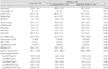
The baseline characteristics of clinical variables and serum collagen markers are shown in Table 1. The age and gender distribution was not different between the three groups. The left ventricular end-diastolic and end-systolic dimensions were significantly lower in the HCM group, especially in the HCM group with a higher PINP/ICTP ratio. The myocardial thickness of the interventricular septum and posterior wall were increased in the HCM group. The HCM group had a significantly larger left ventricular volume index, but there were no differences in the left ventricular systolic function. There were no significant differences in the duration of exercise between the three groups. Of the serum biochemical markers indicating myocardial fibrosis, the log PINP and log PINP/ICTP were significantly higher in the HCM group. Furthermore, the patients with HCM with a higher PINP/ICTP ratio had significantly higher levels of log PINP and log PINP/ICTP due to the study design.
Hemodynamic variables at rest and with exercise
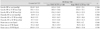
The changes in systolic blood pressure, diastolic blood pressure, and heart rate during exercise are shown in Table 2. In the three groups, the blood pressure and heart rate were increased gradually during exercise; however, there were no significant differences between the three groups in hemodynamic variables at rest and with exercise.
Left ventricular diastolic function at rest
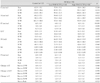
As shown in Table 3, Doppler echocardiographic parameters were compared at rest and each stage of exercise between the three groups. At rest, the E' velocity was significantly lower in the patients with HCM as compared with the control subjects. Interestingly, the HCM group with a higher PINP/ICTP revealed lower E' velocities, a higher E/E' ratio, and lower S' velocities than the HCM group with lower PINP/ICTP. From these results, we know that the left ventricular longitudinal diastolic and systolic function at rest were significantly impaired in patients with HCM who had a higher PINP/ICTP ratio.
Left ventricular diastolic function with exercise
At each stage of exercise, the E' velocity, E/E' ratio, and S' velocity were significantly lower in the HCM group than the control group, and the values were elevated to a similar extent during exercise. Thus, the magnitude of changes in E' velocity, E/E', and S' velocity from rest to each stage of exercise was not different between the three groups. Therefore, the presence of HCM or the higher PINT/ICTP ratio were not associated with a significant decrease in diastolic functional reserve during exercise, even though there was significant left ventricular diastolic dysfunction at rest.
Discussion
The principle findings of this study are as follows: 1) in patients with HCM, the concentration of serum biochemical markers indicating myocardial fibrosis was elevated, and the left ventricular diastolic function at rest was significantly impaired; and 2) although the serum biochemical markers indicating myocardial fibrosis was elevated in patients with HCM, it remained unclear whether the left ventricular diastolic functional reserve was more impaired during exercise.
In patients with HCM, the left ventricular diastolic dysfunction is the most important feature and it has been known as a major determinant of a patient's symptoms.18) The left ventricular diastolic function is determined by the degree of myocardial hypertrophy and interstitial fibrosis. In the normal heart, the intersitial tissue is composed of types I and III collagen. Type I collagen was abundantly present at a ratio of 70 : 30 and it is characterized by a higher degree of rigidity.19) In patients with HCM, due to the presence of myocardial fibrosis, collagen is increased by approximately 20% of the total volume of the myocardium.20) According to this study, type 3 collagen did not show a significant difference in the HCM groups as compared with the control group. Type I collagen and the indicator for the turnover of type I collagen, PINP/CITP in the HCM groups, was higher than the control group. Especially, the patients with HCM with a higher PINP/CITP ratio had a larger septal thickness, decreased diastolic and systolic longitudinal parameters, and increased E over E', indicating a higher left ventricular filling pressure in this group. The differences in these parameters were consistent at rest and at each stage of exercise.
A microscopic examination based on the endomyocardial biopsy has been considered as the most accurate, standard test for the diagnosis of myocardial fibrosis. But, this diagnostic method requires an invasive procedure and there are also limitations that a partial tissue sample of endomyocardium which is obtained from the right ventricle cannot accurately reflect the heterogeneity of myocardial fibrosis in the left ventricle. The type I collagen synthesis-to-degradation ratio, a turnover marker of type I collagen, could estimate the degree of myocardial fibrosis simply with non-invasive serologic tests. Lombardi et al.6) demonstrated a higher collagen turnover in 36 patients with HCM compared with 14 age- and gender-matched controls. In the study, the left ventricular diastolic dysfunction was assessed by the pulse-wave Doppler parameters of mitral inflow and pulmonic vein flow. In the present study, we also assessed the left ventricular longitudinal diastolic function by tissue Doppler imaging. From the tissue velocities on the mitral septal annulus, the E' and S' velocities could be obtained, then the E over E' ratio calculated, indicating the left ventricular filling pressure.
The aim of this study was to prove the hypothesis that a higher level of serologic markers indicating collagen turnover would show a reduced diastolic functional reserve during exercise within the HCM group. However, although the patients with HCM with a higher collagen turnover had significant diastolic dysfunction at rest, there were no significant differences in the degree of changes in diastolic parameters during exercise as compared with the patients with HCM with a lower collagen turnover. Therefore, we conclude that a higher collagen turnover ratio in patients with HCM is not associated with left ventricular diastolic functional reserve in this study population.
The limitations of the current study were as follows: 1) In patients with HCM with a higher collagen turnover marker, there were significantly lower E' and S' velocities from the resting status. Because of these resting differences in the diastolic and systolic tissue parameters, it might be inappropriate to confirm the difference in diastolic function reserve during exercise. To prove the different changes in diastolic function according to exercise, it is preferable that the subjects have similar profiles of E' velocity, S' velocity, and E over E' at rest. 2) This study was limited by the small number of enrolled subjects. 3) This study did not consider the effects of cardiovascular medications which could affect the degree of myocardial fibrosis.
In conclusion, the non-invasive serologic test of type I collagen turnover (the PINP/ICTP ratio) was associated with resting diastolic dysfunction in patients with HCM. However, there was no relationship with augmented diastolic dysfunction during exercise. Therefore, we suggest that the type I collagen synthesis-to-degradation ratio will be a useful marker of resting diastolic function in patients with HCM. Further studies are warranted to examine the diastolic functional reserve with exercise in patients with HCM with relatively well-preserved diastolic function at rest.
 XML Download