

 PDF
PDF ePub
ePub Citation
 Print
Print


Introduction
Significant calcification of coronary artery lesions has been identified as a strong predictor for decreased success and increased complication rates with balloon angioplasty. Calcium imposes a rigid obstacle to optimal and symmetric stent expansion and results in smaller acute gain, and stent under-expansion is associated with an increased risk of acute stent thrombosis and re-stenosis, even with drug-eluting stents (DES). Percutaneous coronary intervention (PCI) for calcified lesions remains a therapeutic challenge. We report a useful technique, the "hugging balloon technique," using two small balloons to treat encircling, heavily-calcified lesions.
Case
Case 1
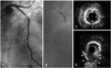
A 76-year-old male patient presented to us with an anterior ST elevation myocardial infarction (STEMI) that developed 3 hours previously. Primary PCI was performed and coronary angiography showed a heavily calcified total occlusion at the proximal left anterior descending (LAD) artery. The lesion was pre-dilated with a 2.5×15 mm balloon (Voyager, Guidant, Indianapolis, IN, USA), but the balloon ruptured at 12 atm and a type C dissection was noted. Two paclitaxeleluting stents (3.0×28 mm, and 3.0×20 mm, Taxus™; Boston Scientific, Boston, MA, USA) were deployed at the midLAD. A follow-up angiogram showed residual stenosis and haziness at the previous calcified total occlusion site (Fig. 1A). After dilatation of the adjuvant balloon 10 times with a 3.0×10 mm balloon (20 atm, Vigor; OCCAM, Eindhoven, Netherlands) and a 3.0×12 mm balloon (Quantum Maverick; Boston Scientific), severe stenosis remained. Intravascular ultrasound (IVUS) revealed an encircling heavy calcification at the previous total occlusion site (Fig. 1C). The hugging balloon technique was performed with two 2.0×15 mm balloons (Splinter; Medtronic, Minneapolis, MN, USA). Two balloons were located at mid-LAD in parallel, and stepwise inflations were performed simultaneously at 12, 18, and 20 atm (Fig. 1B). After the 20 atm ballooning, the lesion site was expanded. Repeated balloon dilatations were performed several times with a 3.0×12 mm balloon (Quantum Maverick). A follow-up angiogram showed resolution of the stenosis and IVUS showed an ellipsoidal rupture of the encircling calcification (Fig. 1D).
Case 2
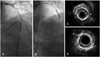
A 68-year-old male was admitted for a STEMI that developed 4 hours previously. Primary angioplasty was planned and the angiogram revealed total occlusion with thrombus and heavy calcification at the mid-LAD artery. After manual aspiration (Thrombuster; Kaneka, Osaka, Japan), an angiogram showed that the heavily-calcified stenosis remained at the mid-LAD artery (Fig. 2A). IVUS revealed a heavy, encircling calcification (Fig. 2C). The lesion was pre-dilated with a 3.0×15 mm balloon (Maverick balloon at 9 atm) and a 3.0×10 mm cutting balloon, but the balloons were not fully dilated at the lesion site. We attempted the hugging balloon technique. Two balloons were located in the mid-LAD artery, side-by-side (2.5×15 mm, and 1.5×15 mm Maverick) and were stepwise inflated simultaneously at 10 and 12 atm. These two balloons were fully expanded and there was no indentation (Fig. 2B). Even though the balloons were fully expanded, the lesion recoiled. However, IVUS revealed breakage of the encircling calcification (Fig. 2D). Then, additional balloon dilation was performed with a cutting balloon. Three sirolimus-eluting stents (2.5×33 mm, 2.75×23 mm, and 3.0×23 mm, Cypher™; Cordis, Johnson & Johnson, FL, USA) were then successfully implanted in the mid-LAD artery. Final angiography showed good dilatation and patency of the LAD artery.
Discussion
These cases highlight the potential efficacy of a double balloon for managing severely calcified lesions. A finding of extensive calcification may lead to the use of plaque modification strategies, such as atherectomy devices or cutting balloon inflation. Rotational atherectomy (RA) has been shown to be efficient for selective calcium ablation.1)2) High-speed RA preferentially cuts hard plaque, increasing plaque compliance, and thereby rendering the lesion more amenable to balloon dilatation.1) However, the potential exists for microcirculatory impairment due to embolization of plaque debris, platelet aggregation, or vasospasm.3) The cutting balloon creates small, sharp, and regular incisions in the plaque and relieves the hoop stress of the vessel, thereby lessening the continuity and rigidity imposed by calcium. The efficacy of the cutting balloon for a calcified lesion has been demonstrated in previous reports,4) but the cutting balloon poses the risk of coronary rupture, microblade fracture and subsequent coronary mural hematoma because
of the blades around the surface of the balloon.5)
A small portion of lesions are refractory to coronary angioplasty, even when very high pressures are used. In double balloon mitral valvulotomy, the tension applied to the plane of the mitral commissures is twice the tension applied vertically, resulting in a more elliptical mitral valve shape, more effective splitting of the fused commissures, and less risk of rupturing or tearing of the leaflets.6) Based on La Place's law for surface tension, higher pressures can be attained with smaller balloons as the burst pressure is higher in smaller balloons compared to larger balloons, as follows: surface tension=pressure×radius. This technique successfully dilates the lesion as the geometry of two balloons inflated side-by-side is different from one balloon. Two balloons consist of two outer semi-circles and a central trapezoidal square area. This altered geometric configuration may be important in successfully dilating a lesion refractory to standard dilating techniques. The determination of adequate diameters is based on the formula, "R2=D12+D22", by adjusting the balloon diameters (D1 and D2) to pre-dict the theoretical mean hugging balloon diameter (R).7)
The preferred choices for management of heavy, calcified coronary lesions are RA or a cutting balloon. However, the judicious use of hugging balloons can be considered in the management of heavy calcified lesions to facilitate adequate dilatation of the lesions when other non-compliant balloons have failed.
 XML Download