

 PDF
PDF ePub
ePub Citation
 Print
Print


Introduction
Left ventricular hypertrophy (LVH) is a major cardiovascular complication and an important predictor of mortality in patients with end stage renal disease (ESRD).1)2)
Wang et al.3) reported that patients undergoing peritoneal dialysis had decreased residual kidney function and cardiac hypertrophy that combined to adversely increase the overall mortality and risk of cardiovascular death.3)
Although echocardiography provides an assessment of LVH and is an important tool used for the evaluation of patients on dialysis, measurement of serum biomarkers that reflect myocyte injury is relatively simple, noninvasive, and may allow better characterization of the nature of cardiac disease when present. Cardiac troponin T (TnT) and N-terminal pro B-type natriuretic peptide (NT-proBNP) have been evaluated as putative prognostic markers of cardiovascular disease in patients with ESRD.4-6) TnT and NT-proBNP have been reported to be correlated with LVH in patients on dialysis.7)8) High sensitivity C-reactive protein (hsCRP), the prototype marker of inflammation, is highly associated with cardiovascular disease in patients with ESRD, and has been reported to be associated with LVH in patients on continuous ambulatory peritoneal dialysis (CAPD).2)
Serum aldosterone levels have been associated with various pathologic patterns of the left ventricle in patients with essential hypertension. In patients with primary hyperaldosteronism, the early onset of LVH and its reversal after treatment is well-known.9)10) The primary role of aldosterone in circulatory homeostasis and salt/water balance is thought to be mediated by the epithelial mineralcorticoid receptor. In addition, aldosterone binds to myocardial mineralocortical receptors and enhances extracellular matrix and collagen deposition in the heart. Aldosterone antagonists have been used in congestive heart failure patients with normal renal function. Some cases have been reported in which the use of spironolactone improves cardiac function without hyperkalemia in peritoneal dialysis patients. Recently, two studies have shown that the left ventricular mass index (LVMI) and serum aldosterone levels are correlated among non-diabetic patients undergoing hemodialysis, and the plasma aldosterone level is independent of blood pressure.11)12)
However, there have been few studies demonstrating a correlation between the LVMI and plasma aldosterone in CAPD patients. We hypothesized that aldosterone could be a useful serum biomarker in predicting LVMI. Because there is a need to determine the best biomarker for predicting LVH, we investigated the relationship between serum biomarkers (TnT, NT-proBNP, CRP, renin, and aldosterone) and LVH in patients on peritoneal dialysis.
Subjects and Methods
Patients
Thirty patients were eligible for the study. The inclusion criteria were continuous peritoneal dialysis treatment for ≥12 months, and no underlying malignancy, chronic liver disease, systemic lupus erythematosus, chronic rheumatic heart disease, or congenital heart disease. All patients had stable body weight and volume status. On physical examination, pretibial pitting edema was not observed and there was no pleural effusion on chest radiography. The heights and weights of all subjects were recorded. The body mass index (BMI) was calculated as the weight (kg)/height (m2).
The blood pressure and body weight were measured before instillation of the dialysate in the peritoneal equilibrium test (PET). The Investigational Review Board of the Cheonan Hospital at Soonchunhyang University approved this study. Informed consent was obtained from all patients who participated in the study.
Biochemical determinations
Blood samples were collected at baseline before PET with patients in the supine position. The plasma was separated after centrifugation at 2,000 g for 15 minutes; the supernatant was stored at -70℃ until used. Routine blood chemistry was performed using standard techniques (76600-020; Hitachi, Japan). Commercially available radioimmunoassay kits were used to measure plasma concentrations of renin (Renin Riabead; Dainabot, Tokyo, Japan), aldosterone (Immunotech SA, France), and intact parathyroid hormone (PTH) (iPTH, PTH-120 MIN IRMA; BioSource Europe SA, Belgium). The hsCRP was measured by a latex high sensitivity assay. The TnT was measured by an electrochemiluminescence assay (Roche modular analyzer; Roche Diagnostic GmbH, Mannheim, Germany).
Echocardiographic measurements
Two-dimensional echocardiography was performed using a Vivid T-dimension (GE, VingMed, Horten, Norway) with a 3.5 MHz probe in subjects lying in the left decubitus position by a single experienced (10 year) echocardiographer blinded to all clinical details about the patients. All findings were confirmed by the cardiologist blinded to all of the patients' clinical details.
Left ventricle (LV) mass was normalized for height2.7 and expressed as the LVMI. The LV mass was obtained by the LV short-axis dimension and a simple geometric cube formula. According to Devereux et al.,13) the following equation provides a reasonable determination of LV mass in grams: 1.04 {(LVID+PWT+IVST)3-LVID3}×0.8+0.6, where left ventricular internal diameter at end-diastole (LVID) represents the internal dimension at diastole, posterior wall thickness (PWT) represents the PWT, interventricular septal thickness (IVST) represents the interventricular septal thickness, 1.04 is the specific gravity of the myocardium, and 0.8 is the correction factor. The height-based indexing of the left ventricular mass (LVM) was specifically chosen to minimize the potential distortion by extracellular volume expansion (body surface area indexing is weightsensitive). An LVMI value >47 g/m2.7 in women and >50 g/m2.7 in men was considered an indication of LVH. The relative wall thickness (RWT) was calculated as PWT×2/LVDd, and a value >0.45 was considered to be increased.14) Using the criteria established by Koren et al.,15) patients with an increased LVMI and increased RWT were considered to have concentric LV hypertrophy, and those with a normal RWT were considered to have eccentric LV hypertrophy.
Pulse Doppler echocardiography was used to assess LV diastolic function. The peak velocity of the early diastolic filling wave (E wave) and atrial filling (A wave) were recorded and the E-to-A ratio (E/A) was calculated. The ventricular filling pressure was estimated by combining the mitral inflow early diastolic velocity (E) and the annulus velocity (E'). The patients were classified according to the E/E' ratio at rest. A value >15 was considered an indication of diastolic dysfunction.
To estimate the left atrium (LA) volume, the biplane area-length formula was used: 8/3π {(A1) (A2)/(L)}, where A1 and A2 represent the maximal planimetered LA area acquired from the apical 4- and 2-chamber views, respectively, and L is length.16) The LA volume index was calculated as the LA volume/body surface area.
Statistical analysis
Data are presented as the mean±SD. A probability value of a p<0.05 was considered statistically significant. All statistical analyses were performed using Statistical Package for Social Science (SPSS) for Windows (version 12.0; SPSS, Inc., Chicago, IL, USA). Continuous variables were analyzed using the Student's t-test, and categorical variables were analyzed using the chi-square test. The Pearson correlation was used to identify the correlation between biomarkers and LVMI.
Results
Patients
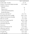
The general characteristics of 30 patients on peritoneal dialysis are summarized in Table 1. Eighteen patients were diabetic, while 12 patients were non-diabetic. Two of the diabetic patients received oral hypoglycemia agents; the other patients received insulin injections. No patients were treated with thiazolinedione. Antihypertensive drugs were used in 30 patients (angiotensin receptor blockers, n=28; calcium channel blockers, n=22; beta-blockers, n=14; minoxidil, n=2). The residual urine volume in 10 patients was <100 mL.
Correlation of aldosterone and the left ventricular mass index
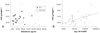
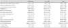
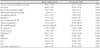
The echocardiography findings are summarized in Table 2. Using the LVMI, 16 of 30 patients had LVH. The serum aldosterone and renin levels were not correlated with the LVMI regardless of the presence of diabetes mellitus (DM) (Fig. 1A); although the plasma aldosterone levels were negatively correlated with the systolic blood pressure (r=-0.524, p=0.003). There was no difference in serum aldosterone levels between patients with LVH and patients without LVH (70.53±76.32 versus 55.83±44.39 pg/mL) (Table 3). The serum aldosterone levels were not correlated with other biomarkers (TnT, NT-proBNP, hsCRP, and renin). The LVMI had a positive correlation with NT-proBNP (r=0.560, p=0.002) (Fig. 1B).
Comparison of the left ventriclular geometry with the biomarkers
According to the LV geometry, nine patients had eccentric LVH (EH), and the other seven patients had concentric LVH (CH). When the EH and CH groups with LVH were evaluated, they had no significant differences associated with the LVMI, renin, aldosterone, or hsCRP. However, NT-proBNP was significantly higher in the CH group than the EH group (5502.31±4301.20 vs. 9797.71±7242.50 pg/mL, p=0.039).
According to the ejection fraction, the LVMI was higher in patients with an ejection fraction <55% compared to the patients with an ejection fraction >55% (39.34±13.11 vs. 23.32±5.22, p<0.01).
Comparison of diastolic markers with the other variables
According to the LA volume index, 16 patients had diastolic dysfunction (≥28 mL/m2). There were no significant differences in serum biomarkers between patients with diastolic dysfunction and those without diastolic dysfunction. The LVMI was significantly higher in patients with diastolic dysfunction (LA volume index ≥28 mL/m2) than in those without diastolic dysfunction (LA volume index <28 mL/m2; 42.48±8.63 vs. 59.12±14.79 g/m2.7, p=0.001). The LA volume index was correlated with the LVMI (r=0.675, p<0.001). According to the E/E', the renin activity was higher in patients with diastolic dysfunction (E/E' ≥15) compared to the other patients (10.76±2.88 vs. 4.67±1.30, p=0.04); however, NT-proBNP was not different between the two groups. The LVMI was significantly higher in patients with diastolic dysfunction (E/E' ≥15) than in those without diastolic dysfunction (E/E' <15; 44.28±10.11 vs. 50.03±16.66 g/m2.7, p=0.025). The E/E' was correlated with the LVMI (r=0.640, p<0.001). The E/A ratio was not correlated with the LVMI.
Discussion
Serum aldosterone has recently been reported to play a significant role in cardiac hypertrophy independent of its effect on blood pressure.17) There are no differences in the mean value of plasma aldosterone between normal subjects and hypertensive patients.18) Aldosterone antagonists have been shown to be effective in patients with heart failure and after an acute myocardial infarction (MI) with adequate renal function. The use of aldosterone antagonists is generally contraindicated in severe renal dysfunction, although some cases have reported that spironolactone could improve cardiac function in ESRD patients. Once patients have progressed to ESRD, aldosterone levels may remain elevated.19) In theory, the elevation of aldosterone might injure cardiac muscle in ESRD patients. In a few studies involving hemodialysis patients, aldosterone was only correlated with the LVMI in non-diabetic patients.11)12) Our study showed that aldosterone was not correlated with the LVMI, even with co-existing DM. We reasoned that this difference could be influenced by dialysis modality and blood pressure.20) In Sato's study,12) a decrease in aldosterone was observed after hemodialysis, possibly due to volume status or clearance by dialysis; however, the volume status is steady in stable peritoneal dialysis. The control of blood pressure might influence the aldosterone level in ESRD. We previously reported that aldosterone concentrations before and after hemodialysis were significantly lower in patients with uncontrolled blood pressure than in patients with well-controlled blood pressure. In the current study, aldosterone was negatively correlated with systolic blood pressure, which is inconsistent with Sato's study.12) Thus, aldosterone levels in CAPD patients might be more influenced by blood pressure than LVMI. In a study of 115 hypertensive patients, Malmqvist et al.21) reported plasma renin activity (PRA) and aldosterone, two markers of the angiotensin-aldosterone system (RAAS), were related to LV mass and RWT, whereas in a study of Korean 275 essential hypertensive patients, PRA was not related to LVMI.22) In our study, PRA was not related to LVMI. RAAS could be influenced by antihypertensive drugs, but in our study, the PRA and aldosterone level were not different according to use of antihypertensive drugs. The results of our study showed that NT-proBNP, among the biomarkers investigated, was correlated with the LVMI. This finding is consistent with previous reports. In our study, troponin T was not correlated with the LVMI and NT-proBNP. NT-proBNP was a superior marker for the prediction of the LVMI compared to TnT in patients receiving peritoneal dialysis. Concentric LVH has been shown to be associated with more marked vascular alterations in ESRD23)24) and a poorer patient outcome.15)25) Our results showed that NT-proBNP and residual renal function might be associated with LV geometry. These results suggest that a worsening of residual renal function might effect the concentric left ventricular remodeling and that NT-proBNP might be a good marker for concentric left ventricular remodeling.
The E/A, E/E', and LA volume are known as markers for diastolic dysfunction. The LVMI has been shown to be associated with diastolic dysfunction.26) The LA volume index has been recognized as a marker of the chronicity of LV diastolic dysfunction.27) In Cho's study,28) the left atrial volume index and the E/E' demonstrated a progressive worsening of the left ventricular diastolic function from patients with normal geometry to the patients with concentric remodeling, and then to the patients with eccentric and concentric hypertrophy in a patient population with hypertension, but without systolic dysfunction. In the present study, the LA volume index and E/E' showed a very good correlation with the LVMI; the LVMI was significantly higher with diastolic dysfunction (LV volume index ≥28 mL/m2 or E/E' ≥15) than without diastolic dysfunction (LV volume index <28 mL/m2 or E/E' <15). Aldosterone was not correlated with diastolic dysfunction marker.
In conclusion, the results of this study suggest that NT-proBNP is a good marker to predict LVH in CAPD patients and aldosterone was not correlated with LVMI, even with co-existing DM in stable CAPD patients.
 XML Download