

 PDF
PDF ePub
ePub Citation
 Print
Print


Introduction
Although drug-eluting stents (DES) significantly reduce restenosis rates compared with bare-metal stents (BMS), treatment of coronary in-stent restenosis (ISR) remains a challenging problem. Much research has been devoted to the pathophysiology and treatment of ISR with DES; however, because of its relatively low incidence, there is scant data on its management.1)
Studies have suggested a variety of possible treatment strategies using standard percutaneous coronary intervention (PCI) techniques, including plain balloon angioplasty (POBA), cutting balloon angioplasty (CBA), repeated DES implantation (Re-DES), radiation therapy, or local drug delivery.2) Some studies reported that repeated PCI with DES, BMS, brachytherapy, POBA or CBA is safe and not associated with increased rates of vascular complications.3-6) Cosgrave et al.7) suggested that Re-DES for DES restenosis is feasible and safe. In particular, sirolimus-eluting stent (SES) failure treated with traditional PCI yielded favorable outcomes, perhaps due to the predominantly focal nature of the SES restenotic lesion.7)8) The use of DES for diffuse ISR is also feasible and safe.9)10)
Regardless of the therapeutic approach chosen, the pattern of restenosis itself is an important predictor of outcomes.7) Similarly, in the era of DES implantation, the incidence of target-lesion revascularization (TLR) increases with the pattern of restenosis treated.11) Therefore, our study was done to evaluate real world outcomes for repeated PCI strategies according to the restenosis pattern in DES failure.
Subjects and Methods
Study population
We reviewed records for 1,465 patients (1,620 lesions) who underwent follow-up coronary angiogram (CAG) from among 2,540 patients who had undergone DES implantation in our 3 centers from April 2003 to March 2006, and identified 83 patients (88 lesions) with DES ISR. Patients who were not treated for ISR or who had left main coronary artery lesions were excluded, and 67 patients (67 lesions) with DES ISR were included in the final analyses. Coronary angiogram follow-up was planned for 9 months after ISR treatment for patients who had not experienced clinical events.
Patients were divided into 3 groups according to restenosis pattern. Group I had focal edge restenosis, Group II had focal body restenosis, and group III had non-focal restenosis. All patients were treated with conventional PCI including POBA, CBA, and Re-DES. All procedures were performed using standard PCI techniques, and intravascular ultrasound (IVUS) imaging was used at the surgeon's discretion. Coronary angiograms were obtained before and after the procedures, and at follow-up, and they were analyzed by 2 independent angiographers. Angiographic and clinical follow-up {major adverse cardiac events (MACEs)} results were evaluated for the 3 groups for 1 year.
Definitions and outcome measures
A focal lesion was defined as one with a length ≤10 mm; non-focal lesions had lengths >10 mm. Edge restenosis was defined as restenosis occurring within 5 mm at either side from and including the stent margin. Angiographic restenosis was defined as diameter stenosis >50% by quantitative coronary angiography (QCA) within a previously stented segment (stent and 5 mm proximal and distal) at follow-up angiogram. TLR was defined as repeated revascularization secondary to a stenosis >50% within the stent or within the 5 mm borders proximal or distal to the stent edge. target-vessel revascularization (TVR) was defined as repeated revascularization of the target vessel. Myocardial infarction (MI) was diagnosed when cardiac creatinine kinase-MB levels were greater than three-fold the normal value, with chest pain lasting ≥30 minutes or with the appearance of new electrocardiographic changes. All deaths were considered to be cardiac-related unless otherwise documented. A MACE was defined as death or MI and the need for TLR, TVR or coronary artery bypass graft (CABG).
Coronary QCAs were analyzed using a validated edge detection system (CAAS II, Pie Medical Imaging, Maastricht, The Netherlands). Minimal luminal diameter (MLD), reference vessel diameter (RD), and % diameter stenosis (DS) were measured at baseline, post-stenting, and at follow-up, respectively.
Statistical analysis
Continuous variables are presented as means±SD, and categorical variables as frequencies and percentages. Differences between groups in outcome variables were assessed using Pearson's Chi-square test or Fisher's exact test (whenever an expected cell value was <5) for categorical data, and the Kruskal-Wallis rank sum test and Wilcoxon rank sum test for continuous data. A p <0.05 was considered to indicate a significant difference, and all reported p are 2-sided. Statistical analysis was performed using SAS 9.1 (SAS Institute, Cary, NC, USA).
Results
Patients and baseline characteristics
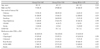
There were 16 focal edge restenotic lesions (group I), 36 focal body restenotic lesions (group II), and 15 non-focal restenotic lesions (group III). Angiographic success was achieved in all patients. Baseline clinical and angiographic characteristics were similar among groups, except for hypertension, which was significantly more frequent in group III (p=0.03). Procedural anticoagulation and antiplatelet therapy was prescribed according to standard protocols. Medications prescribed after DES implantation included aspirin, clopidogrel, cilostazol, beta-blockers, angiotensin converting enzyme inhibitors (ACEI)/angiotensin receptor blockers (ARB), and statins. Frequencies of use of each medication were not significantly different among groups (Table 1).
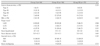
Distribution of target vessels was not significantly different among groups, and the left anterior descending artery (LAD) was the most frequent target vessel. Mean stent length and diameter and proportions by type of inserted stents, i.e., SES or paclitaxel-eluting stent (PES), were not significantly different either (Table 2).
Clinical and angiographic characteristics by percutaneous coronary intervention modality
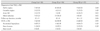
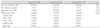
Mean follow-up duration was 10±5 months in group I, 10±8 months in group II and 16±13 months in group III (p=0.06). Acute myocardial infarction frequencies were higher in group III (4, 26.7%) than in group I (2, 12.5%) or in group II (1, 2.8%) (p=0.02), and silent ischemia frequencies were higher in groups I (5, 31.2%) and II (13, 36.1%) than in group III (0, 0.0%) (p=0.02). The latter observation might be related to higher frequencies of routine CAG follow-up in groups I and II than in group III. IVUS studies were done for 27 patients (40.3%), in which neointimal hyperplasia was the major cause of ISR. Stent fracture was seen in 1 (14.3%) case in group I, 8 (57.1%) in group II, and 2 (33.3%) in group III, but the incidence rates were not significantly different (p=0.20) (Table 3).
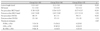
Group III patients had longer mean lesion length and narrower pre-procedure MLD and pre-procedure % DS; however, this was related to grouping characteristics. POBA, CBA and Re-DES were done for the following numbers of patients (and the following percentages by groups): POBA (5, 31.3%), CBA (3, 18.8%), and Re-DES (9, 56.3%) in group I; POBA (13, 36.1%), CBA (17, 47.2%), and Re-DES (6, 16.7%) in group II; and POBA (6, 25.0%), CBA (4, 26.7%), and Re-DES (5, 33.3%) in group III. Frequencies of using treatment strategies were not different among groups (p=0.07) (Table 4).
Angiographic and clinical outcomes at follow-up
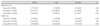
Angiographic follow-up was recommended nine months after repeated PCI with the different therapeutic modalities, and clinical follow-up was planned for 1 year. Anti-platelet medication (aspirin and clopidogrel) coverage was continued in all groups. Angiographic follow-up was performed in 10 (62.5%) patients in group I, 27 (75.0%) patients in group II, and 11 patients (73.3%) in group III (p=0.64); mean follow-up periods were 12±9 months in group I, 10±5 months in group II, and 8±3 months in group III (p=0.33). Restenosis was detected in 3 (30.0%) patients in group I, in 10 (37.0%) in group II, and in 6 (54.5%) in group III, with no statistically significant difference among proportions (p=0.48). A total 19 MACEs developed: 4 (40.0%) in Group I, 6 (22.2%) in Group II and 9 (81.8%) in Group III. The rate of MACEs was significantly higher in group III than in groups I and II (p<0.01) (Table 5).
Comparison of angiographic and clinical outcomes according to therapeutic modality revealed that restenosis rates were not different according to therapeutic modalities within each group. Restenosis rates were as follows: in group I, POBA (1/3, 33.3%), CBA (1/1, 100.0%), and Re-DES (1/6, 16.7%) (p=0.46); in group II, POBA (4/12, 33.3%), CBA (4/10, 40.0%), and Re-DES (2/5, 40.0%) (p=1.00); and in group III, POBA (3/4, 75.0%), CBA (3/4, 75.0%), and Re-DES (0/3, 0.0%) (p=0.14). There was no statistically significant difference in MACE rates between groups I and II. MACE rates were as follows: in group I, POBA (2/3, 66.7%), CBA (1/1, 100.0%), and Re-DES (1/6, 16.7%) (p=0.19); and in group II, POBA (3/12, 25.0%), CBA (2/10, 20.0%), and Re-DES (1/5, 20.0%) (p=1.00). However, in group III, MACE rates were relatively higher for POBA (4/4, 100.0%) and CBA (4/4, 100.0%) than for Re-DES (1/3, 33.3%) (p=0.06), but these differences did not reach statistical significance (Table 6).
Discussion
The main findings of this study are as follows: 1) restenosis rates were not significantly different between focal (edge and body restenosis) and non-focal ISR; 2) restenosis rates were not significantly different among focal edge, focal body and non-focal ISR lesions regardless of the therapeutic modality chosen (POBA, CBA, and Re-DES); and 3) for non-focal ISR lesions, POBA and CBA strategies might be associated with higher MACE rates, and Re-DES might be a better choice for the lesions.
Although ISR after DES implantation is much less frequent than after BMS, it is not less puzzling. Molecular mechanisms of arterial remodeling are less well understood and insight into the mechanisms of DES failure is still limited. It appears that the causes of restenosis after implantation of BMS and DES are fundamentally the same. Restenosis is the arterial wall's healing response to mechanical injury and includes two main processes: neointimal hyperplasia and vessel remodeling.1) Neointimal proliferation is the principal mechanism underlying ISR, and it is the result of endothelial damage after stent expansion.12-14) In our study, IVUS was performed in 27 cases (40.3%), and neointimal hyperplasia was the main cause with stent fracture also being apparent in a total of 11 cases (40.7%). Although some reports have suggested that stent fracture might be another potential risk factor for restenosis, in our study the risk was not significantly elevated; however, there might have been some selection bias, and stent fracture might still be considered as another potential risk factor of ISR.15)16)
The patterns of angiographic restenosis after BMS implantation have been previously described, and it was shown that the Mehran ISR classification is an independent predictor of TLR, emphasizing the prognostic relevance of angiographic features after stent failure. Mehran et al.17) showed that at 1-year follow-up in patients undergoing percutaneous coronary intervention for BMS ISR, a significantly higher rate of TLR occurred with more complex levels of ISR classification. Similarly, in the era of DES implantation, the incidence of TLR increases with the pattern of restenosis treated.11) In the present study, restenosis rates were not different among groups but MACE rates were numerically higher in group III due to the high TLR rate. This result was comparable with that of a previous study.
Currently, there is a paucity of published data on the optimal management of DES restenosis. Some investigators have reported that repeated PCI with DES, BMS, brachytherapy, POBA, or CBA was safe and did not increase rates of vascular complications.2-7)18)19) Most reports on DES restenosis have indicated that the majority of cases, particularly those after SES implantation, are focal. SES failure treated with traditional percutaneous coronary intervention yielded good outcomes at 1-year follow-up (a secondary failure rate of only 23%), perhaps due to the predominantly focal nature of the SES restenotic lesion.8) Systematic use of SES to treat ISR was safe and effective in an unselected series of consecutive patients treated in a real-world scenario, providing very low 9-month ischemia-driven TLR and MACE rates.20) On the basis of BMS-controlled RCT data, there is a definite therapeutic advantage associated with SES and PES use for the prevention of ISR. SES and PES continued to exceed the therapeutic potential of BMS, with a slight but consistent angiographic advantage being observed with SES.4)13)21) Furthermore, the use of DES for diffuse ISR is feasible and safe, and is associated with acceptable early and mid-term results.9)10)20-22) On the other hand, Albiero et al.18) suggested that CBA and traditional PTCA are equally effective in preventing ISR recurrence.
In our study, restenosis rates according to therapeutic modality were not significantly different within each group, and MACE rates were not different between the focal edge and focal body lesion groups. But, even if differences did not reach statistical significance due to relatively small sample sizes, MACE rates were relatively higher for POBA and CBA than for Re-DES for non-focal lesions. Based on our results, it appears that all PCI options are associated with favorable outcomes when used for focal restenotic lesions, while Re-DES appears to be associated with more favorable clinical outcomes for non-focal lesions.
Limitations of this study is that: the data were collected in a retrospective and nonrandomized manner; the sample size was small; there was a relatively low rate of angiographic follow-up. A large scale randomized clinical trial is warranted to determine the optimal strategy for the treatment of ISR after DES implantation.
In conclusion, treatment of DES ISR should be individualized according to restenosis pattern, with all PCI strategies appearing appropriate for focal ISR patterns, and Re-DES appearing to be a better choice for non-focal ISR patterns.
 XML Download