

 PDF
PDF ePub
ePub Citation
 Print
Print


Introduction
Drug-eluting stents have the capacity to reduce neointimal hyperplasia by the local delivery of anti-proliferative agents. While these stents are a very effective solution to the problem of restenosis, restenosis remains a clinical problem.1)2) Recently, stent fracture was suggested as a new potential mechanism of restenosis after sirolimus-eluting stent (SES) implantation.3)4) In this manuscript, we describe stent fracture as a possible cause of restenosis. Several case reports have highlighted the occurrence of stent fracture at follow-up.5) But, few case reports have concerned stent fracture in saphenous vein graft (SVG). We reported the first case of complete SES fracture combined with significant restenosis of a SVG in Korea.
Case
A 74-year-old woman with diabetes mellitus presented with unstable angina in April 2005. She had no other risk factors such as hypertension, hypercholesterolemia, alcohol, or smoking. She revealed a diagnosis of three-vessel coronary disease upon coronary angiography, and underwent coronary artery bypass graft surgery with placement of a SVG to the right coronary artery and SVG to the left anterior descending artery.
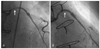
Two years later, she was admitted with unstable angina. Coronary angiography revealed 70% focal stenosis of the proximal SVG to the left anterior descending artery (Fig. 1A). The SVG to the left anterior descending artery was predilated with a 2.0×15 mm angioplasty balloon at 6 atm. After predilation, a 2.75×18 mm Cypher® SES (Cordis, Roden, The Netherlands) was deployed in the SVG and inflated at 10 atm (Fig. 1B). She tolerated the procedure well and was discharged without chest pain.
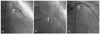
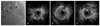
A follow-up coronary angiography was performed after 6 months. On admission to the hospital, the patient displayed a pulse rate of 68 beats/min, blood pressure of 110/70 mmHg and respiratory rate of 18 breaths/min. Her physical examination was essentially normal. A chest X-ray demonstrated no abnormalities except sutured metallic rings around the sternum. A complete blood count, electrolytes and thyroid function tests were within normal limits. Cardiac enzymes were also within normal limits. An electrocardiogram showed normal sinus rhythm without ST-segment change. Transthoracic echocardiogram revealed good regional wall motion with ejection fraction of 69%. The coronary angiography revealed 80% focal in-stent restenosis with complete stent fracture (Fig. 2A and B). Intravascular ultrasound showed neointimal hyperplasia at mid-stent without presence of a stent strut (Fig. 3). The stenosis was crossed with a guide wire and dilated with a 3.0×15 mm angioplasty balloon at 20 atm. A final coronary angiography showed thrombolysis in myocardial infarction (TIMI) 3 distal flow of the SVG without residual stenosis (Fig. 2C).
Discussion
In-stent restenosis after implantation of SESs still occurs in some cases, and stent fracture was recently suggested as a new potential mechanism of restenosis.3)4) The "real world" clinical practice of implantation of SESs may be a safe and feasible technique for revascularization in patients with SVG disease with excellent short- and long-term clinical outcomes.6) But, the use of bare-metal stents may be associated with lower long-term mortality than the use of SES for SVG disease.7) An incidence of stent fracture after SES implantation of 2.6% was reported.8) While the exact mechanism of stent fracture is unknown, stent fractures are associated with long stented segments, right coronary artery location and metal overlap.9) Multiple mechanical factors such as long stented segments, increased rigidity and pre-procedure vessel angulation might interact to disrupt the stent strut. The majority of stent fractures occur within 10 mm from areas of increased rigidity caused by strut overlap that may have acted as a fulcrum for metal deformation due to vessel movement.
Dynamic vessel movement and repetitive kinking of the stent during the cardiac cycle may also be implicated in stent fractures.9) Many drug-eluting stent fractures occur in the proximal segments of the right coronary artery, since this artery moves more dynamically than the left coronary artery during the cardiac cycle.10)
The implantation of multiple overlapping stents significantly increases the axial stiffness of the stented segment and longer stents covering longer lesions are subject to higher radial forces. These may play a role in stent fracture.9) SES fracture may be due to the closed cell design of the SES compared with the paclitaxel-eluting stent, which has an open cell design.11)
In-stent restenosis after implantation of a SES is commonly associated with a discontinuity in stent coverage.12) Late incomplete stent strut coverage because of stent fracture could result in incomplete inhibition of intimal hyperplasia and subsequently in restenosis at follow-up.10) Therefore, fractured stent struts cause a local mechanical irritation of the vessel and may result in inflammation and neointimal hyperplasia.11) At the fracture point, neointimal growth is putatively observed because of the decrease in local drug delivery.8) Furthermore, exposure of a free metal strut protruding into the vessel lumen clearly could trigger platelet activation and resultant stent thrombosis.11)
In our case, a short stent was deployed in the proximal curvature portion of SVG to the left anterior descending artery. Unlike the native coronary arteries, the body of a SVG is relatively free to move in relation to the anastomotic site, depending upon the degree of perigraft fibrosis and amount of intrathoracic space available. Consequently, in vein grafts, mechanical stresses can be very high.4)13)
As yet, there is no consensus regarding the best treatment method of stent fracture. Re-stent therapy is controversial, since there is a possibility of recurrence of stent fracture. Overlapping stents may also increase the likelihood of stent fractures from increased axial stiffness at the overlapping segment acting as a fulcrum for stent deformation and fracture from vessel movement.14) Furthermore, the effect of balloon angioplasty for uncovered SES restenosis is unknown.8) Instead of re-stent therapy, we treated stent fracture with balloon angioplasty. Follow-up studies should be performed to confirm that there is no in-stent restenosis.
Stent fracture of the SVG is relatively rare. The presently-reported case is the first case of complete stent fracture of SVG in Korea.
 XML Download