

 PDF
PDF ePub
ePub Citation
 Print
Print


Sinoatrial node (SAN) automaticity is responsible for controlling heart rhythm. SAN function is therefore essential for normal cardiac physiology. Although it was shown that spontaneous diastolic depolarization of SAN cells periodically initiates action potentials to set the rhythm of the heart, the mechanism by which heart rhythm is generated is unclear. Sick sinus syndrome is an abnormality involving the generation of the action potential by the SAN and is characterized by an atrial rate inappropriate for physiologic requirements. The sick sinus syndrome occurs in 1 of every 600 cardiac patients with >65 years of age and accounts for approximately one-half of implantations of pacemakers in the United States.1) A better understanding of the mechanisms of SAN automaticity is therefore clinically important.
The Voltage Clock and Sinoatrial Node Automaticity
The mechanism of spontaneous diastolic depolarization has traditionally been attributed to a "voltage clock" mechanism, mediated by voltage-sensitive membrane currents, such as the hyperpolarization-activated pacemaker current (If) regulated by cyclic adenosine monophosphate (cAMP).2)3) This current is also referred to as a "funny" current because, unlike the majority of voltage-sensitive currents, it is activated by hyperpolarization rather than depolarization. The funny channel becomes activated at the end of the action potential (at voltages from -40/-50 mV to -100/-110 mV), which corresponds to the range in which diastolic depolarization occurs. The funny channel then depolarizes the membrane to a level at which Na+ channels open to initiate the action potential.4)5) If is a mixed Na+-K+ inward current activated by hyperpolarization and modulated by the autonomic nervous system. Because the membrane ionic channels open and close according to the membrane potential, this process is referred to as a membrane voltage clock. The f-channels are encoded by the hyperpolarization-activated, cyclic nucleotide-gated (HCN) channel gene family. Of the four known HCN subunits, HCN4 is the most highly expressed in the mammalian SAN. The major role of If has been reinforced by the fact that mutations in the If channel are associated with a reduced baseline heart rate,6) and drugs which block If (such as ivabradine) do the same.7)8) However, while point mutations of HCN4 are associated with baseline sinus bradycardia, the maximum heart rate achieved during exercise is normal.9) The latter finding implies that If is not the only mechanism of SAN automaticity, especially during sympathetic activation.
The Calcium Clock, a New Mechanism for Sinoatrial Node Automaticity
The traditional mechanism for heart rhythm generation has been restricted to sarcolemmal ion channels. Recently, spontaneous Ca2+ release from the sarcoplasmic reticulum (SR) has been suggested to be the mechanism for sinus rhythm generation. When the SR is full, the probability of spontaneous Ca2+ release increases. On the other hand, when the SR is empty, the chances for spontaneous Ca2+ release decreases. The rhythmic alterations of SR Ca2+ release is referred to as the Ca2+ clock. Because the SR Ca2+ content is controlled in part by the membrane voltage, it is important to recognize that the activation of the Ca2+ clock and the membrane ionic clock are interdependent. The involvement of Cai in heart rhythm generation was first suggested by the observation that application of ryanodine slowed subsidiary pacemakers in cat right atria.10) Subsequent work showed the involvement of the electrogenic Na-Ca exchange current (INCX) in pacemaker activity and also raised the possibility that increased Ca2+ release, followed by activation of INCX, could play a role in the positive chronotropic effect of β-adrenergic stimulation in latent pacemaker cells.11-16) Lakatta et al. suggested that spontaneous rhythmic Ca2+ release from the SR in SAN cells, manifested as Ca2+ sparks, work as the "Ca2+ clock."17-21) The elevated Cai causes diastolic depolarization via INCX activation, which coordinately regulates the sinus rate along with the voltage clock.17-21)
The idea of a Ca2+ clock suggests that the mechanism of automaticity is the same as that of delayed afterdepolarization (DAD), which occurs when there is a SR Ca2+ overload. This suggests that the SAN must exist normally in a state of calcium overload. Vinogradova et al.21) also demonstrated that a high basal level of protein kinase A (PKA) activity in the SAN might contribute to a state of Ca2+ overload. A disease associated with Ca2+ clock malfunction has also been reported in patients with a genomic deletion of the RyR2 exon-3. Patients with that mutation develop catecholaminergic polymorphic ventricular tachycardia, along with SAN and atrioventricular node dysfunction, atrial fibrillation, and atrial standstill.22) It is possible that Ca2+ clock malfunction might contribute to the bradycardia and atrial arrhythmias in the latter patients.
Pacemaker Hierarchy and the Importance of the Ca2+ Clock in an Intact Sinoatrial Node
Cardiac automaticity at the organ level is a very complex phenomenon and, in addition to cellular mechanisms, integrative factors are involved in cardiac pacemaking. The intact SAN is a heterogeneous structure that includes multiple different cell types interacting with each other.23-26) The relative importance of the voltage and Ca2+ clocks for pacemaking in different regions of the SAN, and in response to neurohumeral stimuli, such as β-agonists, may be different. Indeed, activation maps in intact canine right atria (RA) have shown that the SAN impulse origin is multicentric,27) and sympathetic stimulation predictably results in a cranial (superior) shift of the pacemaking site in humans and dogs.28)29) Based on evidence from isolated SAN myocytes, late diastolic Cai elevation (LDCAE) relative to the action potential upstroke is a key signature of pacemaking by the Ca2+ clock.10-21) It is possible that the same phenomenon could provide insight into the relative importance of the Ca2+ and voltage clock mechanisms in pacemaking in intact SAN tissue. We therefore designed a study to determine the role of Ca2+ and voltage clocks on heart rhythm generation and on the mechanisms of pacemaker hierarchy in the intact SAN.30)
Heterogeneous Cai Dynamics in an Intact Sinoatrial Node
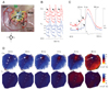
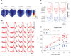
At baseline, the spontaneous diastolic SR Ca2+ release, which is manifested by the LDCAE, was observed in only a small percentage of the preparations. However, the LDCAE occurred in all preparations during isoproterenol infusion, and was associated with a superior shift of the leading pacemaker site coincident with the appearance of robust LDCAEs (Fig. 1) in this region. Most importantly, the site of maximum LDCAE slope always co-localized with the leading pacemaking site, suggesting a shift in which the voltage clock now lagged the Ca2+ clock (Fig. 2). This observation indicates a strong association between LDCAE and pacemaking during β-adrenergic stimulation, and provides new insight into pacemaker hierarchy in the canine RA.27-29)
The Cai dynamics of SAN were characterized not only by the earliest onset of LDCAE, but also by the fastest Cai reuptake as compared with other RA sites. The baseline 90% Cai relaxation time was shorter at the superior SAN than at other RA sites. This resulted in the formation of the Cai sinkhole, which was facilitated by a rapid decline (short relaxation time) of the Cai fluorescence at the superior SAN during isoproterenol infusion and suggests that Cai reuptake by the SR is fastest in the superior SAN (Fig. 1D). The key protein regulator of SR Ca2+ uptake is phospholamban, which inhibits SERCA2a in the dephosphorylated state. There was a significantly lower SERCA2a/phospholamban ratio at SAN sites than at RA sites, suggesting more phospholamban molecules are available to regulate SERCA2a molecules in SAN than in RA. Isoproterenol infusion phosphorylates phospholamban and relieves phospholamban inhibition of SERCA2a, which may account for more robust Ca2+ uptake in SAN than in RA during isoproterenol infusion.30)
The Importance of Sarcoplasmic Reticulum Function on Pacemaking
The LDCAE was closely related to SR Ca2+ release. Caffeine sensitizes the ryanodine receptor 2 to activation, resulting in increased SR Ca2+ release.18) The superior shift of the LDCAE and the pacemaking site was also consistently observed with caffeine infusion. Ryanodine, which blocks ryanodine receptors, caused a dose-dependent suppression of sinus node activity, and impaired isoproterenol-induced LDCAE.
The combination of ryanodine and thapsigargin also suppressed sinus node activity, and impaired isoproterenol-induced LDCAE. In contrast, the If blocker, ZD 7288 (3 µmol/L), did not prevent the LDCAE in the superior SAN.
Mechanisms of Diastolic Depolarization
Multiple time-and voltage-dependent ionic currents have been identified in cardiac pacemaker cells, which contribute to diastolic depolarization, including ICa-L, ICa-T, IST, and various types of delayed rectifier K currents.31) Many of these membrane currents are known to respond to β-adrenergic stimulation. Some of these currents, such as ICa-L, also promote the LDCAE and the acceleration of the sinus rate by the Ca2+ clock, as well as the voltage clock. In an intact SAN, SR inhibitors and If blockade slowed the sinus rate under basal conditions and blunted the isoproterenol-induced increase in the sinus rate. Therefore, the interdependence and synergy between the two clocks was evident.
Sympathetic Stimulation and Tachybrady Syndrome
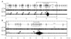
It is known that heart failure is frequently associated with SAN remodeling, resulting in decreased SAN reserve.32) We performed nerve recording in a canine model of pacing-induced heart failure and found intermittent tachybradycardia episodes.33) Interestingly, the prolonged (>3 s) sinus pauses were triggered not by vagal activation, but by short bursts of sympathetic activity (Fig. 3). Typically, a burst of sympathetic activity is associated with tachycardia. When there is sympathetic withdrawal, the tachycardia terminates, followed by prolonged pauses during which no activation was observed. Preliminary studies34) showed that cryoablation of the stellate ganglia markedly reduces the prolonged sinus pause episodes in the same model. These findings suggest that the left stellate ganglion nerve activity is causally related to the tachybrady syndrome. The molecular mechanism of this association, however, remains unclear.
Future Directions
Our understanding of cardiac automaticity has progressed considerably during the last 10 years. Several questions regarding how the heart rhythm is generated and controlled in physiologic and pathologic conditions remain unanswered. A recent study by Yeh et al.35) showed funny current downregulation is assocciated with atrial tachyarrhythmias. The authors suggested that the down-regulation of funny current may underlie the mechanisms tachybrady syndrome. Whether or not Ca2+ clock malfunction also contributes to the mechanisms of tachybrady syndrome remains unclear. Our laboratory is currently evaluating whether the Ca2+ clock is impaired in a canine atrial fibrillation model with sinus dysfunction. Secondarily, we are trying to reproduce the human sick sinus syndrome model by manipulating the Ca2+ and voltage clocks in a canine isolated RA preparation. We anticipate that these studies will help us better understand the mechanisms of SAN dysfunction and facilitate design mechanism-based therapy of this disease.
 XML Download