

 PDF
PDF ePub
ePub Citation
 Print
Print


Introduction
Thrombi causing a pulmonary thromboembolism (PTE) mostly involve the peripheral veins, and in particular, the central veins of the lower extremities; a PTE also may occur in the pelvic veins. Accordingly, a PTE can be regarded as a complication of a venous thromboembolism (VTE), and thus the risk factors of VTE, and in particular the risk factors of deep vein thrombosis (DVT), are considered the risk factors of a PTE. A renal venous thrombosis (RVT) as the origin of a PTE is very rare in relatively healthy individuals without thrombogenic diseases.1)
Oral contraceptives (OCs) are the most common method of hormonal contraception. In general, OCs have been shown to be safe for most women, and in addition to the effect of contraception, OCs have advantages, such as an increase in bone density, a decrease in menstrual blood loss, a decrease in ectopic pregnancies, and a reduced risk of ovarian cancer. Rarely, however, OCs cause DVTs and PTEs in users.2)
Case
A 28-year-old woman presented with a chief complaint of syncope. She had a feeling of congestion in her chest and flank for approximately 1 week, and on the day of admission to the hospital, she collapsed in the restroom and lost consciousness for a few seconds, and thus she was referred to the emergency room immediately. She was previously healthy except for having a termination of an ectopic pregnancy with methotrexate about 4 months previously. The only medication she was taking was an OC, which was started 3 months before admission. Diane 35 (cyproterone acetate 2 mg/ethinyl estradiol 0.035 mg; Bayer Schering Pharma AG™) was used as the OC initially; however, it was changed to Mercilon (desogestrel 0.15 mg/ethinyl estradiol 0.02 mg; N.V. Organon™) due to urticaria after taking Diane 35 for 1 week. She had no family history of a VTE. On examination, she was appeared acutely ill. Her consciousness was clear, and the body temperature, blood pressure, and pulse rate were 36℃, 110/70 mmHg, and 78/minute, respectively. The respiratory rate increased to 28/minute. She weighed 63 kg and was 167 cm tall {body mass index (BMI), 22.59 kg/m2}. The heart sounds were regular and no cardiac murmurs were appreciated. The lungs were symmetric and the breath sounds were clear. The liver, spleen, and kidneys were not palpable, and there was no edema, stasis, or varices in the lower extremities.
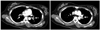
On admission, the chest X-ray showed mild cardiomegaly, and an electrocardiography (ECG) demonstrated a regular sinus rhythm with a right axis deviation. The white blood cell count was 8,800/mm3, the hemoglobin was 12.7 g/dL, the hematocrit was 38.1% and the platelet count was 138,000/mm3. The results of the urine, liver function, renal function, and electrolyte tests were all normal. An arterial blood gas analysis on room air was as follows: pH, 7.48; PaCO2, 26.4 mmHg; PaO2, 48.0 mmHg; bicarbonate, 19.2 mmol/L; oxygen saturation, 87.4%; and P(A-a)O2, 61.0 mmHg. The prothrombin time was 1.05 international normalized ratio (INR) and the activated partial thromboplastin time was 23.6 seconds. The N-terminal pro-B-type natriuretic peptide (NT pro-BNP) was 1,154 pg/mL. A 2D echocardiogram revealed normal left ventricular systolic function and dimension, with a slightly flattened interventricular septum and an enlarged right atrium and ventricle, and the estimated pulmonary artery pressure was 42.6 mmHg. Because a PTE was suspected, a CT scan was performed, including the chest to the lower extremities. The CT scan showed that a massive PTE in the distal part of the pulmonary artery, the interlobular artery, and the segmental artery on both sides, and right ventricular hypertrophy was noted (Fig. 1). Moreover, a huge RVT was observed (Fig. 2A). However, there was no evidence of a DVT in the lower extremities on CT venography. Venous ultrasonography did not demonstrate venous thrombosis involving the lower extremities. Under the diagnosis of an acute PTE, the patient received thrombolytic therapy (tissue-type plasminogen activator, 50 mg for 1 hour and 50 mg for 2 hours) followed by intravenous heparin, and was closely monitored in the intensive care unit. Her chest discomfort improved dramatically following thrombolytic therapy. Five days after thrombolytic therapy, a 2D echocardiographic assessment revealed significant reduction of the right ventricular dimension and a normalized pulmonary arterial pressure.
In assessment of intrinsic thrombogenecity, it was determined that she had no evidence of other known risk factors. The serum titers of anticardiolipin antibody (IgG and IgM) were within normal limits (1.2 GPL and 1.7 MPL U/mL, respectively). Antithrombin III (78.5%), and protein C and S activities were normal (1.45 and 0.30 mg/dL, respectively). Lupus anticoagulant, and antinuclear and antineutrophil cytoplasmic autoantibodies (ANCA) were negative. The homocysteine was 7.0 umol/L and Factor V Leiden was G/G.
The patient was continued on intravenous heparin and an oral anticoagulant (coumadin) during the admission (7 days). She was discharged with an INR of approximately 2.5. The PTE had resolved completely on the follow-up CT scan obtained on day 20 of systemic anticoagulants. The RVT remained on the follow-up CT on day 20 (Fig. 2B), but resolved completely by day 50 (Fig. 2C). Warfarin was discontinued 3 months later. One year after this event, she was pregnant and healthy.
Discussion
The clinical symptoms of VTE and PTE are nonspecific and many cases do not show any symptoms. For this reason, the diagnosis of PTE is more difficult than treatment or prevention, and the frequency and mortality rates are not certain. The most useful approach is the clinical assessment of the likelihood of a PTE, based on presenting symptoms and signs, in conjunction with judicious diagnostic testing. Syncope is a relatively rare presenting symptom of a PTE, but portends a life-threatening event when associated with a PTE.3) However, this patient had other symptoms suspicious for PTE, including chest discomfort, dyspnea, and tachypnea. In cases of a massive PTE, as with this patient, immediate 2D echocardiography is a very helpful diagnostic tool for further evaluation. Considering the increased tendency, acute PTE should be included in the differential diagnosis in evaluating patients who present with syncope.3) According to reports, there are approximately 5 million new cases of DVTs in the US every year, 500,000 (10%) of which develop into a PTE, and leading to 50,000 deaths.4) In >95% of the cases, thrombi occurs in the deep veins of the lower extremities, and thrombi involving the deep veins above the knee often cause a PTE. Thrombi may occur in the pelvic veins, the veins of the upper extremities, the hepatic veins, the renal vein, and the right atrium.1)
The risk factors for PTE and VTE include a previous history of a VTE, heart disease, cancer, trauma, major surgery, pregnancy, exogenous estrogen use, paralysis or profound immobility, and increasing age. Therefore, it is important to evaluate the underlying causes and origin of the thrombus rather than the diagnosis of the PTE only.1) In this regard, chest CT scanning for suspected PTE should include CT venography of any vein that might contain thrombi. Indeed, some chest CT imaging protocols for suspected PTE routinely image the abdomen, pelvis, thighs, and knees.5) As in this case, we also have a similar CT imaging protocol for evaluating PTE and this was very helpful for evaluating the originating site of the thrombi. In this case, there were no other thrombogenic risks, except OC, and a history of an ectopic pregnancy.
A VTE is the most common vascular disease in women in their reproductive years. In women in their reproductive years who do not use OCs, VTEs occur at a rate of around 1/10,000. Since OCs were first introduced in the 1960s, venous thrombi have increased in young women and this increase is known to be closely related to the dosage of estrogen.6) The administration of high-dose estrogen resulting from the use of OCs increases coagulation factors V, VIII, and X, and the production of fibrinogen.7) As OCs with a low concentration of estrogen have been developed, currently used OC regimens contain <50 µg of estrogen, but Sidney et al.8) reported that OCs with low estrogen content still increase the risk of VTE by 3-4 times, and the risk is highest in the first year of the use.
In 1995, there were controversies triggered by the report that among low-dose oral contraceptives, the 3rd generation OCs containing gestoden and desogestrel had twice the risk in causing VTE than 2nd generation OCs containing levonorgestrel.9) On the other hand, Suissa et al.10) conducted a comparative study on the period and pattern of use of OCs in women who began to use 2nd and 3rd generation OCs and reported that there was no corresponding difference in the frequency of VTEs. Based on 5 years of comparative research, Lidegaard et al.2) reported that the risk of VTE decreases over time in women who are currently using OCs, while OCs containing desogestrel or gestodone increase the risk of venous thrombosis slightly, and the risk of a VTE is slightly higher in women who smoke 10 or more cigarettes per day. In addition, they reported that the risk of a VTE is lower in OCs containing 20 µg of estrogen than in OCs containing 30-40 µg, and contraceptives containing only progesterone do not increase the risk of a VTE. The patient reported herein used a 3rd generation OC containing 150 µg of desogestrel and 20 µg of estrogen. The occupation of this patient was a telemarketing counselor. She worked about 10 hours a day during the last 4 years without any exercise. A sedentary lifestyle, a history of an ectopic pregnancy, and OC use might cause an increase in hemostasis and thrombogenicity in such patients.
RVT usually presents as an insidious, chronic disease in patients with nephrotic syndrome or renal cell carcinoma. Acute RVT is rare in adults and usually occurs after blunt abdominal trauma or renal transplantation.1)2) The use of OCs has been considered as a relatively weak risk factor for RVT compared to DVT.
Few cases have been reported, but there are no reports involving a PTE associated with a RVT due to OC use alone in a patient without any combined underlying thrombogenic diseases. Thus, we have reported a case of a woman who had a PTE and a RVT while taking OCs with a review of the relevant literature.

 XML Download