

 PDF
PDF ePub
ePub Citation
 Print
Print


Introduction
The objective of cardiac pacing is to optimize cardiac performance. This is dependent on three main parameters: the chronotropic function of the sinus node or pacemaker, the quality of atrioventricular synchrony across the spectrum of rates and global inotropic states as governed by autonomic tone, and the intraventricular activation sequence. The advent of intravenous lead delivery systems established the right ventricular apex (RVA) as the standard site for pacemaker lead implantation. However, several studies have demonstrated the deleterious consequences of RVA pacing in patients with cardiomyopathy.1)2) Abnormal conduction of the paced depolarization through the ventricular myocardium results in dyssynchronous left ventricular (LV) contraction due to slow intramuscular conduction, in comparison to brisk conduction through the His-Purkinje system.3) The net result is impaired systolic and diastolic function. Furthermore, LV dyssynchrony may cause myocardial perfusion abnormalities that further diminish cardiovascular efficiency and function.4)5)
These observations have led to an interest in selecting new RV pacing sites in order to achieve a more physiological pattern of ventricular activation.6)7) Therefore, other sites have been proposed for RV pacing lead implantation: the RV outflow tract (RVOT),8)9) RV free wall,10) and His bundle area.11) The ventricular activation sequence observed in RVOT pacing may more closely resemble that occurring during normal His-Purkinje activation. We hypothesized that RVOT pacing is associated with less mechanical dyssynchrony compared to RVA pacing and that it more closely resembles mechanical activation in normal control patients with a narrow QRS.
Subjects and Methods
Patient population
We enrolled 9 patients who underwent radiofrequency catheter ablation for supraventricular tachycardia (SVT) and who had normal LV systolic function and no regional wall motion abnormalities noted during echocardiography performed before the procedure; these subjects served as the study group. We also enrolled 9 patients with idiopathic left bundle branch block (LBBB) with a QRS duration of >120 msec. Fifteen normal subjects were included as controls. All patients gave informed consent before enrollment in the study.
Study design
Nine patients were brought to the EP lab for electrophysiologic study and ablation of SVT. After completing the radiofrequency catheter ablation for SVT, pacing was performed via a temporary pacing catheter (Biosense Webster, Diamond Bar, CA, USA, 2-5 mm interval) positioned under fluoroscopic guidance in the RVA and then in the RVOT. Pacing was performed at each site at threshold with a cycle length of 500 msec for a duration of 3 minutes. Echocardiographic myocardial images were stored with color coding at the end of each pacing train. The time interval between pacing trains was 10 minutes. Echocardiographic myocardial images were also obtained in two sets of controls, including patients with idiopathic LBBB and patients with normal QRS duration. We evaluated and compared the degree of LV contraction and dyssynchrony in four groups of patients: 1. RVA pacing, 2. RVOT pacing, 3. patients with LBBB in normal sinus rhythm (NSR), and 4. patients with normal QRS in NSR. We excluded the patients in whom we could not measure the Doppler parameters in stored images. In all patients, the QRS width was measured as the widest among all the leads.
Echocardiography
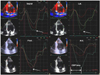
Each individual underwent a full echocardiographic examination before and after the procedure using a Vingmed Vivid 7 (General Electric, Horten, Norway) system. The images were stored digitally on a commercially available software system (Echopac 6.3.6; GE Vingmed, Horten, Norway) and analyzed offline. The echocardiograms were analyzed in pairs with the reporter blinded to the data of each scan. The LV ejection fraction was assessed using Simpson's biplane equation for calculating volumes. The LV diameter and mass were measured in the parasternal long axis view using M-mode. Tissue Doppler color imaging was performed using the standard apical 4- and 2-chamber views for the long-axis motion of the LV with a frame rate of over 100/sec. At least 3 consecutive beats in each view were stored and digitized for offline analysis. The mean data of three beats was used for the final analysis. From the apical 4- and 2-chamber views, four different LV wall segments-namely the septal, lateral, inferior, and anterior segments-were selected, and then three sample volumes of 5 mm were positioned at the basal, middle, and apical segments in each wall (Fig. 1). The myocardial strain profile was reconstituted from the tissue Doppler color images. The echocardiographic examination was performed with the same method in the resting state in the normal and LBBB groups.
The myocardial systolic strain was measured at the compression/expansion crossover point (CEP) (Fig. 1), which represented the degree of actual contraction of the LV. We also measured the time from the QRS onset to the CEP, which was defined as the CEP time. Since this CEP time represents the exact time-point of the actual contraction time of each segment, it is a useful tool for analyzing dyssynchrony.12)13) The degree of LV dyssynchrony was represented by the dispersion and standard deviation (SD) of the CEP times. The dispersion was obtained by calculating the difference between the maximum and minimum CEP times from all studied segments. In addition, the dispersion was corrected for the heart rate (HR) using the Bazett formula14) {corrected dispersion (msec½)=dispersion (msec)/HR (msec)½}, because the dispersion could be affected by the heart rate. The inter- and intra-observer concordances of CEP time were 88% and 92%, respectively.
Statistical analysis
The SPSS 12.0 (SPSS, Inc., Chicago, Illinois) statistical software package was used for all calculations. Data are presented as means±SDs for continuous variables and as percentages for the categorical data. Differences between groups were analyzed using the analysis of variance (ANOVA) test. Categorical data were analyzed using the Chi-square test. P<0.05 were regarded as statistically significant.
Results
QRS width
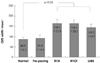
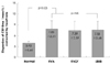
The QRS duration of normal and pre-pacing beats was significantly shorter than that for the RVOT and RVA pacing and LBBB groups. There was no significant difference in QRS duration among the RVA pacing, RVOT pacing, and LBBB groups (Fig. 2).
Degree of left ventricular contraction
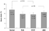
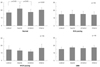
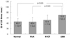
The mean strain was similar for the pacing and LBBB groups and was significantly lower than that seen in the normal control group (Fig. 3). In terms of sub-analysis of the strain in each wall among the groups, the strain in the septal wall in the normal group was significantly higher. However, in the pacing and LBBB groups, we could not find the prominent area of strain (Fig. 4).
Degree of left ventricular dyssynchrony
In terms of the changes in the total contraction time, the mean CEP time corrected for heart rate was similar between the RV pacing and LBBB groups and was significantly higher than that in the normal control group (Table 2). When the dispersion of the CEP time was corrected for the heart rate, the difference between the RV pacing and LBBB groups nearly disappeared; the dispersion of the CEP time in the aforementioned groups was significantly higher than that in the normal group (Fig. 5). In addition, the standard deviation of the CEP time in the LBBB group was significantly increased compared to those in the normal and RVA pacing groups. However, there was no statistically significant difference in SD between the normal and RV pacing groups (Fig. 6).
Discussion
The principal finding in this study was that the degree of regional myocardial contraction and dyssynchrony assessed by Doppler tissue strain imaging was similar in the RVA and RVOT pacing and LBBB groups. Based on these findings, RVOT pacing may not be a more favorable alternative and may result in LV dysfunction similar to that observed in patients with RVA pacing or those with LBBB.
Several authors have reported conflicting findings concerning the acute and chronic differences in hemodynamic effects between RVA and RVOT pacing.15-19) A pooled analysis of nine prospective studies evaluating the hemodynamic effects of RVOT pacing in 217 patients indicated a modest but significant hemodynamic benefit compared with RVA pacing.15) Giudici et al.16) reported that the cardiac output improved at the time of implantation with RVOT pacing, from 6.6 L/min at the apex to 7.8 L/min at the outflow tract, a 19% increase. In contrast, Buckingham et al.18) demonstrated an insignificant increase in the dP/dt during single site RVOT pacing, as compared to single site RVA pacing. Victor et al.19) found neither a substantial change in the functional class nor a hemodynamic benefit during RVOT pacing as compared to RVA pacing during midterm follow-up.
There are two possible explanations for these mixed results. First, the position of the lead in the RVOT could have influenced the results. Most of the previous studies described the lead position using X-ray images, and only some of them identified the final lead position using the pacing-induced QRS width.15)17) The different investigators paced at different sites in the RVOT. Tse17) and Stambler et al.20) reported that the QRS width in RVOT pacing was shorter compared to that in RVA pacing. However, there was a different QRS width for RVOT pacing in the two studies (134 ms vs. 167 ms, respectively). Increasing distance of the RV pacing site from the His-Purkinje system causes prolongation of the QRS interval and greater intramyocardial conduction time, which may provoke LV dyssynchrony.21) Riedlbauchova et al.22) suggested that the ideal pacing position seems to be the mid-septum, where the earliest endocardial signal can be observed, often with a potential from the right bundle. This was reflected by a narrow QRS complex as compared to RVOT pacing. In our study, we positioned the pacing lead in the high septum using fluoroscopic and ECG morphologic guidance, involving a LBBB and inferior axis. However, the QRS width for RVOT pacing was 163 ms.
Second, pacing-induced acute changes in hemodynamic performance do not necessarily predict hemodynamic improvement during long-term pacing. Like the adverse hemodynamic and clinical effects of spontaneous LBBB, the iatrogenic variety of the LBBB produced by conventional RVA pacing technique employed in permanent cardiac pacing may be equally harmful.23) Our study showed a similar degree of contraction, assessed by Doppler tissue strain imaging, among the RVOT pacing, RVA pacing, and LBBB groups. This suggests that RVOT pacing-induced LBBB exhibited similar contractile dysfunction compared to that seen in the idiopathic LBBB group.
In the present study, we measured the dispersion of the CEP time in the 12 segments using Doppler tissue strain imaging as the index of dyssynchrony. Patients with heart failure have concurrent electrical delay on the surface ECG, mainly in the form of LBBB. This ventricular conduction disturbance changes the LV contraction pattern, resulting in dyssynchronized intra- and inter-ventricular contraction and further impairing systolic performance. In addition, abnormal myocardial shearing forces and stress vectors may manifest as abnormal contractility and dyssynchrony.24) Doppler tissue imaging, strain rate, and strain have been demonstrated to be useful tools in evaluating myocardial dyssynchrony.25-27) Doppler tissue strain imaging can measure the local myocardial shortening and lengthening and is less sensitive to segment tethering and translation than is tissue Doppler imaging.28) The strain image may be used as a more precise tool for quantifying ventricular contractility and dyssynchrony.
There are a few limitations to this study. We chose a 3-minute pacing period. It is possible that the results may have been different if a longer period had been allowed for conditions to be established. However, two papers,18)29) which measured echocardiographic and hemodynamic parameters after pacing by using catheters at different sites, also used 2- or 3-minute pacing protocols. Additionally, a relatively small number of patients was analyzed in this study. A large-scale, longterm follow-up study will be required in the future. Another limitation of our study is that dyssynchrony was measured using only Doppler tissue strain imaging instead of strain rate imaging. Acute findings in dyssynchrony and regional contraction may be related to chronic adverse responses. However, it is unclear if these acute findings predict long-term clinical effects.
In conclusion, the present study suggests that RVOT pacing, which is characterized by a wide QRS width, is associated with significant mechanical dyssynchrony and is not a viable alternative to RV apical pacing. As a result, RVOT pacing may have an adverse effect on long-term LV function similar to that observed with RV apical pacing.


 XML Download