Abstract
Late stent thrombosis is one of the most serious complications associated with morbidity and mortality after coronary drug-eluting stent implantation, and is mainly caused by the withdrawal of antiplatelet agents. We report our experience of late stent thrombosis simultaneously involving three different coronary arteries in a young male patient who was treated with three drug-eluting stents two years ago. The patient stopped taking antiplatelet agents for several days. The patient did not recover from cardiogenic shock, even after repeated ballooning with thrombus aspiration, intra-aortic balloon pumping, and temporary pacing during cardiopulmonary resuscitation.
Go to : 
References
1. Grube E, Silber S, Hauptmann KE, et al. Two year-plus follow up of paclitaxel-eluting stent in de novo coronary narrowing (TAXUS I). Am J Cardiol. 2005; 96:79–82.
2. Degertekin M, Serruys PW, Foley DP, et al. Persistent inhibition of neointimal hyperplasia after sirolimus-eluting stent implantation: longterm (up to 2 years) clinical, angiographic, and intravascular ultrasound follow-up. Circulation. 2002; 106:1610–3.
3. Fajadet J, Wijns W, Laarman GJ, et al. Randomized, double-blind, multicenter study of the Endeavor zotarolimus-eluting phosphorylcholine-encapsulated stent for treatment of native coronary artery lesions: clinical and angiographic results of the ENDEAVOR II trial. Circulation. 2006; 114:798–806.
4. Moreno R, Fernandez C, Hermandez R, et al. Drug-eluting stent thrombosis: result from a pooled analysis including 10 randomized studies. J Am Coll Cardiol. 2005; 45:954–9.
5. Schampaert E, Moses JW, Schofer J, et al. Sirolimus-eluting stents at two years: a pooled analysis of SIRIUS, E-SIRIUS, and C-SIRIUS with emphasis on late revascularizations and stent thrombosis. Am J Cardiol. 2006; 98:36–41.
6. Iakovou I, Schmodt T, Bonizzoni E, et al. Incidence, predictors, and outcome of thrombosis after successful implantation of drug-eluting stent. JAMA. 2005; 293:2126–30.
7. Daemen J, Wenaweser P, Tsuchida K, et al. Early and late coronary stent thrombosis of sirolimus-eluting and paclitaxel-eluting stents in routine clinical practice: data from a large two-institutional cohort study. Lancet. 2007; 369:667–78.

8. Park SH, Hong GR, Seo HS, Tahk SJ. Stent thrombosis after successful drug-eluting stent implantation. Korean Circ J. 2005; 35:163–71.

9. McFadden EP, Stabile E, Regar E, et al. Late thrombosis in drug-eluting coronary stents after discontinuation of antiplatelet therapy. Lancet. 2004; 364:1519–21.

10. Moon JY, Jeong MH, Jun CH, et al. The frequency, treatment and clinical outcomes of stent thrombosis after use of TAXUS stent. Korean Circ J. 2007; 37:641–6.

11. Wernick MH, Jeremias A, Carrozza JP. Drug-eluting stents and stent thrombosis: a cause for concern? Coron Artery Dis. 2006; 17:661–5.

12. Park DW, Park SW. Stent thrombosis in the era of the drug-eluting stent. Korean Circ J. 2005; 35:791–4.

13. Ong AT, Hoye A, Aoki J, et al. Thirty-day incidence and six-month clinical outcome of thrombotic stent occlusion after bare-metal, sirolimus, or paclitaxel stent implantation. J Am Coll Cardiol. 2005; 45:947–53.

14. Ong AT, McFadden EP, Regar E, de Jaegere PP, van Domburg RT, Serruys PW. Late angiograhpic stent thrombosis (LAST) events with drug-eluting stents. J Am Coll Cardiol. 2005; 45:2088–92.
15. Joner M, Finn AV, Farb A, et al. Pathology of drug-eluting stents in humans: delayed healing and late thrombotic risk. J Am Coll Cardiol. 2006; 48:193–202.
16. Collet JP, Montalescot G, Blanchet B, et al. Impact of prior use or recent withdrawal of oral antiplatelet agents on acute coronary syndromes. Circulation. 2004; 110:2361–7.

17. Goel PK, Gill GS. Angioplasty for the late stent thrombosis, the new technical challenge: a single centre experience. Int J Cardiol. 2008; 123:335–7.

18. Burger W, Chemnitius JM, Kneissl GD, Rucker G. Low-dose aspirin for secondary cardiovascular prevention-cardiovascular risks after its perioperative withdrawal versus bleeding risks with its continuation: review and metaanalysis. J Intern Med. 2005; 257:399–414.
Go to : 
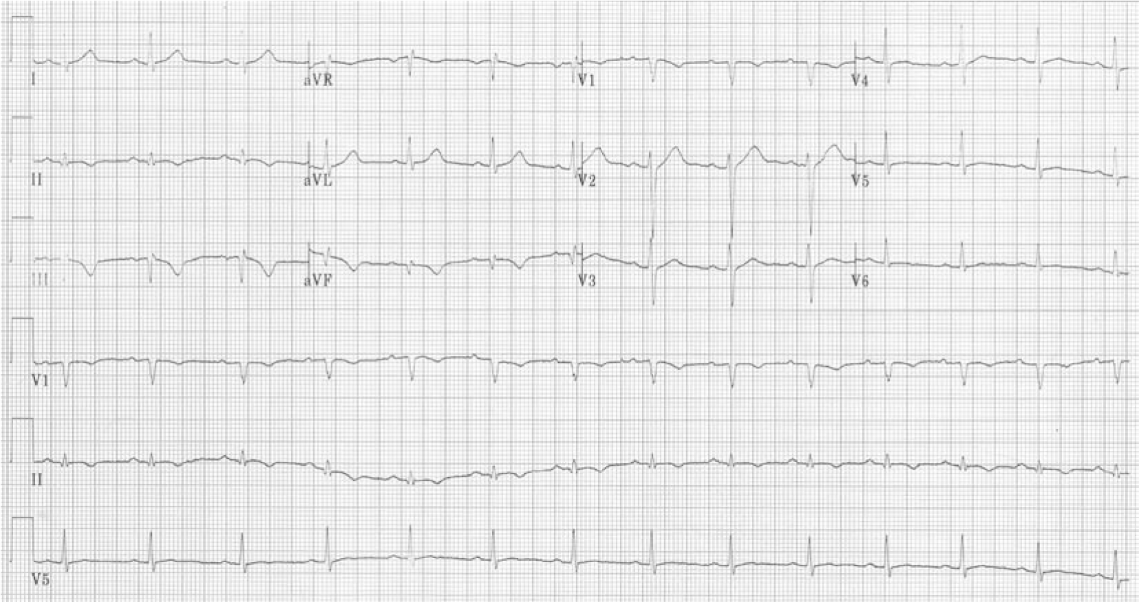 | Fig. 1.The first cardiac event, ECG obtained in the emergency room of another hospital showed Q and T wave inversions in the inferior leads. ECG: electrocardiogram. |
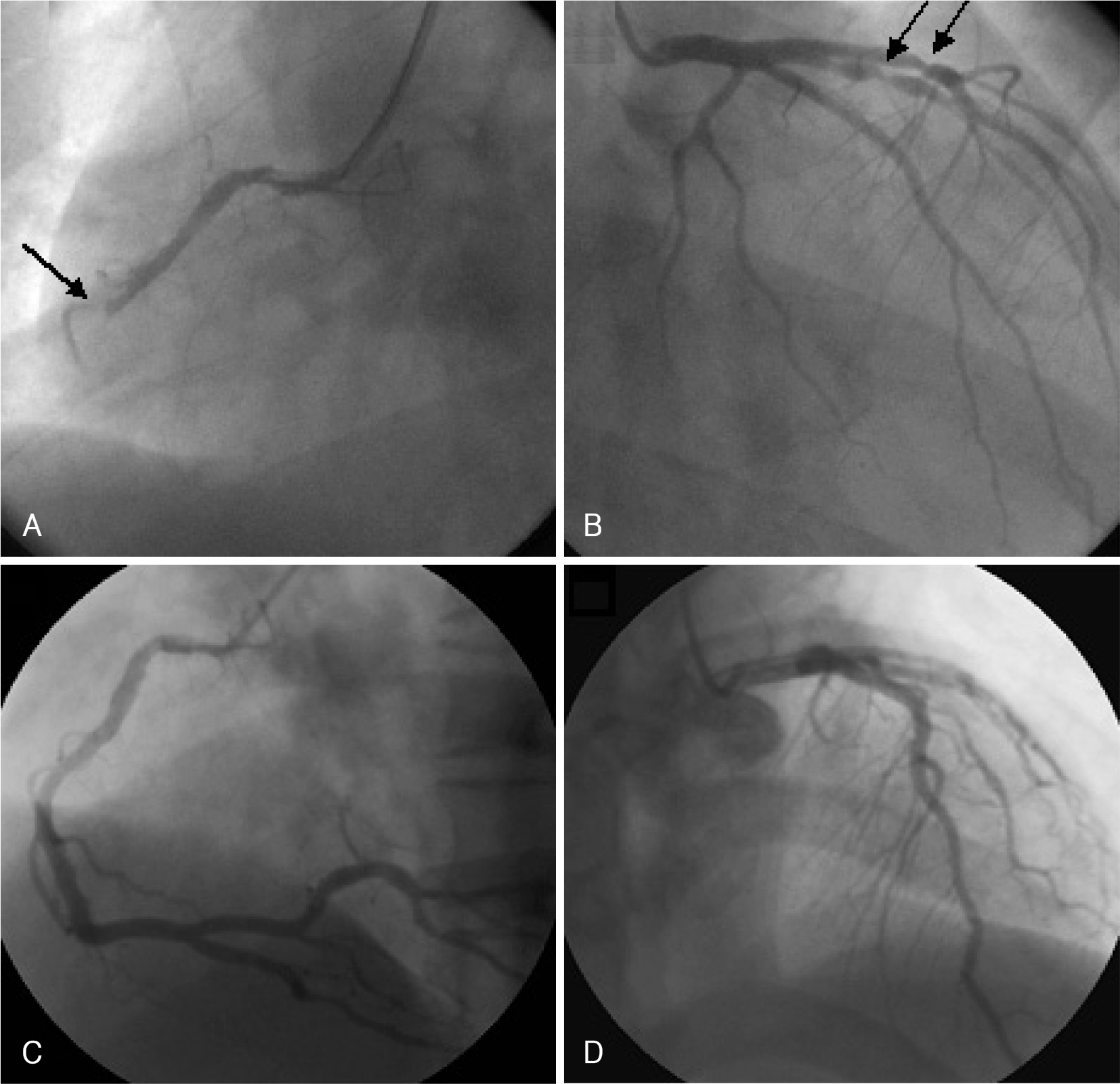 | Fig. 2.During the first cardiac event. A and B: an emergency coronary angiography revealed total occlusion of the middle RCA, and critical stenosis in the proximal LAD and intermediate branch (A: LAO view, B: RAO cranial view). C and D: after coronary stenting, distal flow was restored to TIMI III in three coronary arteries (C: LAO view, D: AP cranial view). RCA: right coronary artery, LAD: left anterior descending artery, LAO: left anterior oblique, RAO: right anterior oblique, AP: anterioposterior, TIMI: thrombolysis in myocardial infarction. |
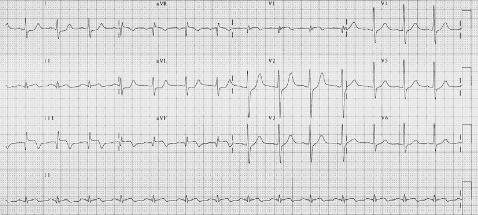 | Fig. 3.The ECG taken in the emergency room during the second cardiac event showed ST elevation in the inferior leads. ECG: electrocardiogram. |
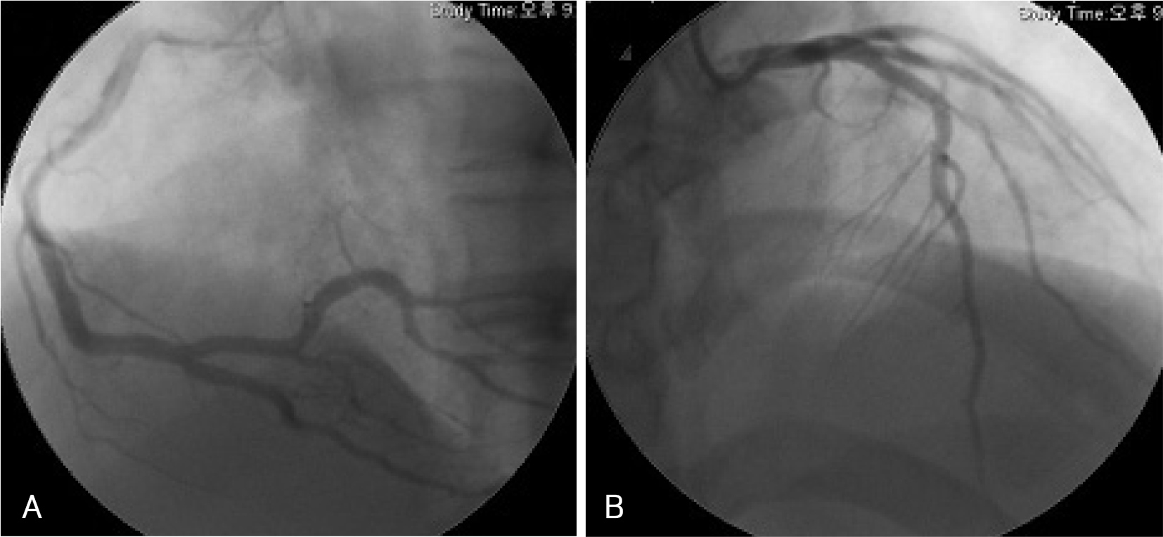 | Fig. 4.During the second cardiac event (A and B) an emergency coronary angiography revealed patency in the previous stented sites ofthe middle right coronary artery, the proximal left anterior descending artery, and the intermediate branch with good distal flow (A: LAO view, B: AP cranial view). LAO: left anterior oblique, AP: anterioposterior. |
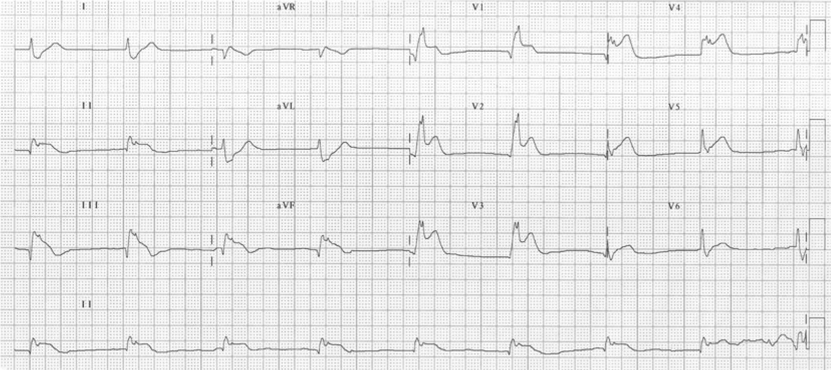 | Fig. 5.The ECG obtained in the emergency room during the third cardiac event revealed complete AV block, a right bundle branch block, and ST elevation over the precordial and inferior leads. ECG: electrocardiogram, AV: atrioventricular. |
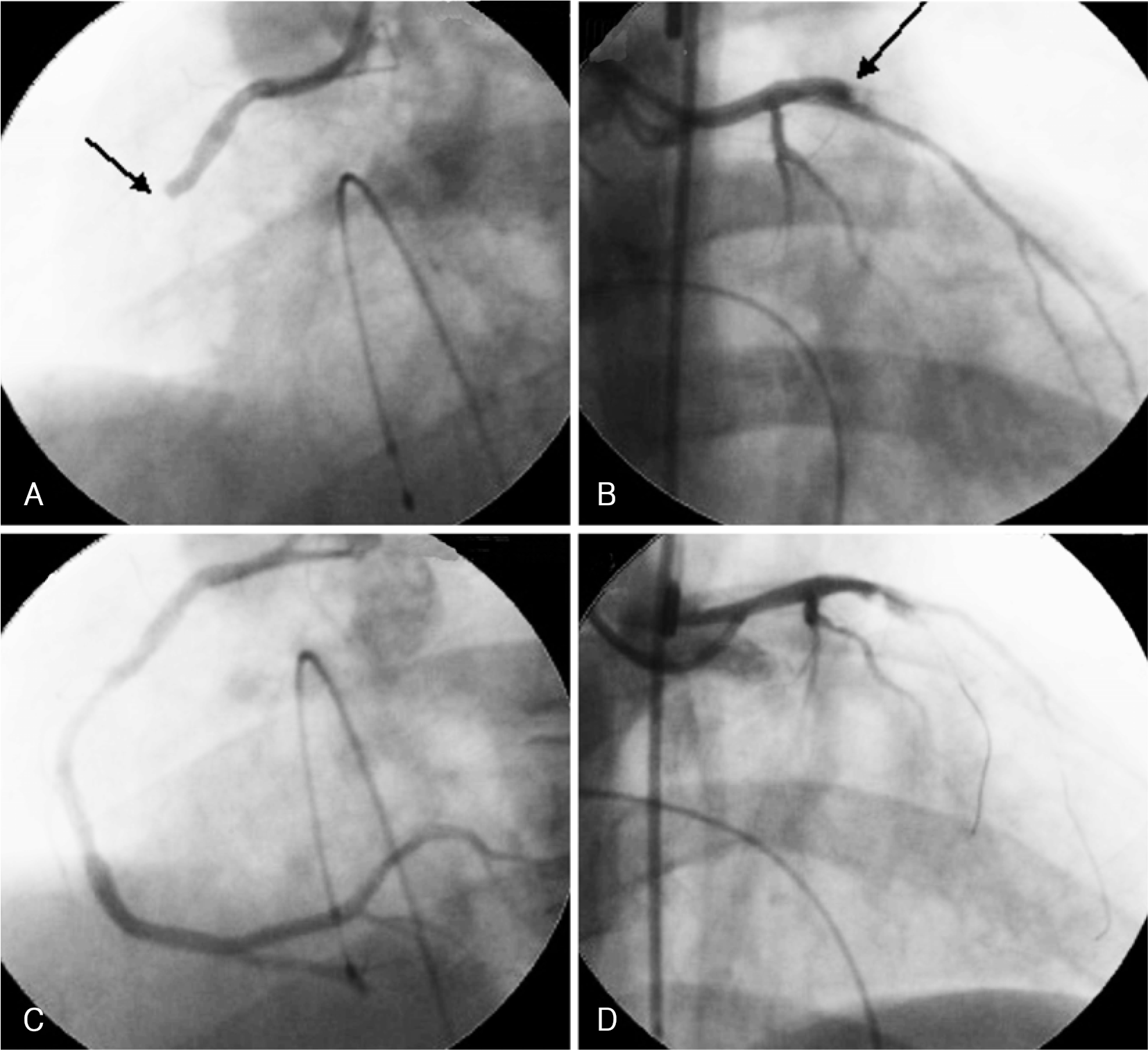 | Fig. 6.During the third cardiac event. A and B: an emergent coronary angiography revealed simultaneous total occlusion of the middle RCA, proximal LAD, and intermediate branch, at the sites the three Taxus stents had been previously placed (A: LAO view, B: RAO cranial view). C and D: after placement of an intra-aortic balloon counterpulsation and temporary pacemaker, thrombus aspiration was performed by suction catheter. But, coronary flows at the occluded lesions of the proximal LAD and intermediate branch were not restored in spite of repeated ballooning and thrombus aspiration (C: LAO view, D: RAO cranial view). RCA: right coronary artery, LAD: left anterior descending artery, LAO: left anterior oblique, RAO: right anterior oblique. |


 PDF
PDF ePub
ePub Citation
 Print
Print


 XML Download