

 PDF
PDF ePub
ePub Citation
 Print
Print


Introduction
Pulmonary arterial hypertension (PAH) is a fatal disease that's caused by progressive narrowing of the pulmonary arterioles and increased pulmonary vascular resistance, and this can result in right ventricular failure and death.1)2) There are several factors that play roles in the pathogenesis of PAH such as genetic mutation of bone morphogenetic protein receptor II (BMPR II) and activin receptor-like kinase 1 (ALK-1), exposure to drugs, infection and inflammation. These factors eventually lead to multiple abnormalities, including endothelial cell proliferation, matrix production, thrombosis, abnormal vascular vasoconstriction and vascular remodeling.2)
The currently available treatments for PAH include adjunctive therapy like diuretics, anticoagulant, digoxin and oxygen for right heart failure, and pulmonary vasodilating agents such as calcium-channel blockers, prostanoids and endothelin receptor antagonists.3) However, these currently available therapies can not reverse the disease process or the considerable morbidity and mortality, although they have a beneficial clinical effect.4) Therefore, new therapies are warranted that are based on a better understanding of the pathophysiology of the disease, and especially those therapies that are directed at suppressing inappropriate cellular proliferation in the pulmonary arteries.2)5)
Simvastatin is a well known drug with cardiovascular benefits that exceed their effects on lowering serum cholesterol. In addition, simvastatin also exert potent antiproliferative and proapoptotic effects on vascular smooth muscle cells through the inhibiting the activities of ras and rho guanine triphosphatase (GTPase), which are important for cell proliferation.6) It has recently been demonstrated that simvastatin could reverse established, severe pulmonary hypertension after toxic injury in rats and improve their survival. Other studies also showed that simvastatin treatment potently attenuates chronic hypoxic pulmonary hypertension in rats and it inhibits vascular remodeling.2)7-10)
The aquaporins (AQPs) are a family of small membrane-spanning proteins that are expressed on the plasma membranes of many cell types and they facilitate water transport. In the lung, AQP1 is expressed on the apical and basolateral membranes of the microvascular endothelial cells. The abundance of AQP1 in the peribronchiolar vessels of perinatal rats may suggest their role in clearance of lung water from the alveolar space.11) Renal AQP2 is present in the vesicles of the collecting duct principal cells that translocate to the apical membrane in response to vasopressin.12) AQP2 overexpression has been described in several conditions associated with fluid retention, including congestive heart failure.13) These results suggest that AQP may play a role in the edema formation in PAH. Several animal models of PAH revealed pulmonary microvascular leakage and interstitial inflammation, which resulted in pulmonary edema.14) The aims of this study were examine the protective effect of simvastatin on monocrotaline (MCT)-induced PAH and the role of the AQP water channels.
Materials and Methods
Experimental designs
Twenty one male Sprague Dawley rats (180-200 g) were randomly divided into three groups: group 1 was injected with distillated water (the control group, n=7), group 2 was treated with MCT (the MCT group: MCT was injected at a single dose 60 mg/kg, sc, n=7) and group 3 was MCT rats that were treated with simvastatin (Hanmi Pharmacy, Seoul, Korea) (the MCT+S group, 5 mg/kg/day, po, daily for 28 days from the onset of MCT treatment, n=7). The rats in all three groups were kept in the same room and all of them were subjected to the same light-dark cycle. After 28 days, their tissue samples were obtained for morphometric analysis and western blotting. The experimental procedures we used were reviewed and approved by the Animal Care and Use Committee of Dongguk University. The animal care and use were in accordance with the guidelines of the National Institute of Health.
Measurement of right ventricular pressure
After 28 days, the animals were anaesthetized by an intraperitonial injection of ketamine (100 mg/kg). For measuring the right ventricular pressure, a PE 50 catheter (Becton Dickinson, Franklin Lakes, NJ, USA) was inserted into the right ventricle via the internal jugular vein, and a fluid-filled PE-50 tube was connected to a pressure transducer (Grass polygraph, Grass instrument CO, Quincy, MA, USA).
Right ventricular hypertrophy and lung morphology
After 4 weeks, the rats were euthanized by an anesthetic overdose and then the right ventricle (RV) free wall was dissected from the left ventricle (LV) and septum (S), and they were weighed separately on the analytic scale. The RV remodeling was assessed by the RV-to-LV plus S weight ratio. For analyzing the vascular remodeling, the left lung was fixed with a transcardiac infusion of 4% paraformaldehyde. The perfused lung was removed and then paraffin-embedded. Serial coronal sections 5 µm thick were obtained at the lower zone of the lung. Following deparaffinization, the sections were stained with the hematoxylin-eosin (H&E). The medial wall thickness (MWT) of the pulmonary arterioles was measured at the pulmonary arterioles that were 50-100 µm in size and at the peribronchiolar muscular arteries. The MWT ratio, which is an index of medial wall hypertrophy, was determined as the average data of 10 to 15 fields per slice and the MWT ratio was calculated as: [MWT=(external diameter-internal diameter)/external diameter].
Western blotting of aquaporin1 and aquaporin2
The MCT and/or simvastatin treated lung and kidney tissues were removed and then snap-frozen at -70℃ for Western Blot analysis. The tissue samples were homogenized in ten volumes of homogenizing buffer (0.32 M sucrose, 25 mM imidazole and 1 mM ethylenediaminetetraacetic acid (EDTA) (pH 7.2) containing 8.5 mM leupeptin and 1 mM phenylmethylsulfonyl fluoride), for 10 s with using a polytron. The aliquots were stored at -70℃. Samples of the homogenate were run on 7.5% polyacrylamide mini gels (Bio-rad Mini Protean). For each gel, an identical gel was run in parallel and the two gels subjected to Coomassie staining to assure identical loading. After electrophoresis, the protein was transferred to nitrocellulose paper for 2 hours at 400 mA and 120 V in a BioRad transblot system. After transfer, the protein bands were identified by Ponceaus S and they were destained with distilled water. The nitrocelluse sheets were washed in tween phosphate buffered saline (PBST) and then incubated with rabbit anti-AQP1 (Alomone, Jerusalem, Israel) and rabbit anti-AQP2 (Alomone, Jerusalem, Israel) for overnight at 4℃. The labeling was visualized with horseradish peroxidase-conjugated secondary antibodies (Santa Cruz Biotechnology, Santa Cruz, CA, USA) and with using an enhanced chemiluminescence (ECL) system (Amersham Pharmacia Biotech, Little Chalfont, UK). The immunoblot signal was developed by an ECL system and it was quantified using Scion Image software (version 1.59).
Laboratory findings
On day 26 after treatments, the animals were individually housed in metabolic cages. The daily dietary intake and urine volume were measured for 2 days. The rats' blood and urine samples were stored for electrolyte and renal function testing.
Statistical analysis
All the data is presented as means±SDs. The data was analyzed by one-way analysis of variance (ANOVA) followed by Tukey's multiple-comparisons test. Multiple-comparisons tests were applied only when a significant difference was determined by ANOVA (p<0.05). P<0.05 were considered statistically significant.
Results
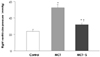
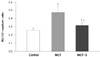
The effect of simvastatin on the high right ventricular pressure and right ventricular hypertrophy in rats with monocrotaline-induced pulmonary hypertension
The right ventricular pressure was significantly increased in the MCT group as compared with that of the controls (52.4±3.9 vs. 24.1±0.26 mmHg, respectively, p<0.05), and the right ventricular pressure was markedly suppressed in the simvastatin treatment group (32.3±2.1 mmHg, p<0.05). However, simvastatin treatment did not suppress the right ventricular pressure to normal values (p<0.05) (Fig. 1).
In the MCT group, right ventricular hypertrophy developed and there were significant increases in the RV/LV+S ratio compared with that of the controls (0.48±0.07 vs. 0.26±0.02, respectively, p<0.05), and the right ventricular pressure was markedly suppressed in the simvastatin treatment group (0.32±0.03, p<0.05). However, simvastatin treatment did not completely suppress the RV hypertrophy to normal values (p<0.05) (Fig. 2).
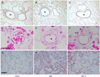
The effect of simvastatin on the wall thickness of the pulmonary artery in the rats with monocrotaline-Induced pulmonary hypertension
MCT treatment increased the medial wall thickness (MWT ratio) of the peribronchiolar artery compared with that of the controls (0.28±0.02 vs. 0.10±0.03, respectively, p<0.05), and the medial wall thickness was significantly suppressed in the simvastatin treated group (0.14±0.02, p<0.05) (Fig. 3A-C). The medial wall thickness of the small pulmonary arterioles (50-100 µm) was also significantly reduced in the simvastatin treated group as compared with the MCT group (0.15±0.04 vs. 0.29±0.11, respectively, p<0.05) (Fig. 3D-F). This treatment, however, did not completely reverse the pulmonary arterial wall thickness to a normal value (p<0.05). Perivascular and interstitial edema was clearly seen in the MCT group. However, simvastatin treatment significantly reduced the pulmonary edema (Fig. 3G-I).
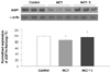
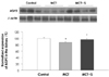
The effect of simvastatin on lung aquaporin1 and renal aquaporin2 in the rats with monocrotaline-induced pulmonary edema
To quantitatively evaluate the effect of simvastatin on MCT-induced pulmonary hypertension, the AQP1 and AQP2 expressions were measured in the lung and kidney from the controls and the MCT and/or simvastatin treated rats. Western blot analysis demonstrated that the expression of lung AQP1 and renal AQP2 in the MCT group was significantly decreased compared with that of the control group (p<0.05). However, the expression of lung AQP1 and kidney AQP2 was normalized after treatment with simvastatin (p<0.05) (Figs. 4 and 5).
Laboratory findings
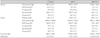
The serum osmolarity and sodium levels were slightly increased in the MCT group (p<0.05), but they were normalized after simvastatin treatment (p<0.05). Other parameters of renal functions such as the urine output and the Na, K, blood urea nitrogen, creatinine and creatinine clearance were not altered (Table 1).
Discussion
PAH is a disease that's characterized by elevated pulmonary artery pressure, and this can lead to right ventricular failure and death.1)2) The median survival of PAH patients was reported to be just a few years with an estimated 5-year survival of 34%.15-17) Yet the newer medical therapies, besides the adjunctive therapies, have been shown to improve a variety of clinically relevant end-points, including survival, exercise tolerance, the haemodynamics and the quality of life measures.2)5) The introduction of continuous intravenous prostacyclin, a dual endothelin receptor antagonist, for the treatment of PAH has been promising.3) Unfortunately, there is no curative therapy. Therefore, the current therapy for PAH is based on a better understanding of its pathogenesis and an effort to reverse the vascular remodeling with using phosphodiesterase type 5 inhibitors, elastase inhibitor, platelet derived growth factor (PDGF) receptor antagonist and simvastatin.12)18)19)
Simvastatin improves the cardiovascular outcomes, and this is independent of its effects on cholesterol reduction. In addition to its potent antipoliferative and antiapoptotic effects on the vascular smooth muscle cells,20) simvastatin enhances the production of endothelial nitric oxide synthase (NOS)21) and it also has anti-inflammatory effects.22) Simvastatin treatment causes activation of caspase-3 and pulmonary microvascular endothelial cell apoptosis in cases of severe pulmonary hypertension.10) It has recently been demonstrated that simvastatin could reverse or attenuate pulmonary hypertension.9)23) A small study reported improvements of the 6 min walk tests and the hemodynamics in patients who received simvastatin daily.6) Clinically, we showed that simvastatin decreased the right ventricular pressure, the right ventricular hypertrophy and the medial wall thickness of the pulmonary artery in the rats with MCT-induced PAH. These results suggest that simvastatin attenuates the progression of MCT-induced pulmonary hypertension through the reduction of vascular resistance. This study was not intended to explore the mechanisms of simvastatin's effects, but the increased apoptosis of vascular smooth muscle cells and/or the endothelial cells and production of endothelial NOS are might be candidate mechanisms that prevent the increase of vascular resistance.
The existence of water-specific membrane channel proteins in selected tissues has been postulated for several decades, and eleven mammalian AQPs have now been identified.12) The abundance of AQP1 in the peribronchiolar vessels may suggest a role for this protein in clearance of water from the interstitial space.11) In ventilator-induced lung injury, AQP water channels have protective effects on pulmonary edema formation.24) However, in a murine model of lipopolysaccharide-induced acute lung injury, depletion of AQP1 did not affect the formation of lung edema, the lung vascular permeability or the lung histology.25) These results suggest that any correlation between the decreased expression of AQP1 and lung edema formation is controversial. In this study, we revealed that the APQ1 expression was decreased in the MCT-induced PAH rat tissues and this was normalized with simvastatin treatment. The APQ1 expression was also correlated with the resolution of lung edema. Recent papers have reported that AQP depletion would have subclinical effects on water homeostasis, and these effects would become apparent under stressful conditions such as congestive heart failure and pulmonary edema.12)26) Therefore, we suggest that simvastatin might attenuate the formation of pulmonary edema through the decreased expression of AQP1 water channels.
In a previous rat model of congestive heart failure caused by acute myocardial infarction, the renal AQP2 expression was activated in the apical plasma membrane of the renal collecting ducts, which accounted for fluid overload.13) Further, the up-regulation of APQ2 was normalized after fosinopril, valsartan and losartan treatment.27)28) So, we hypothesized that the fluid retention with pulmonary edema in PAH would be related with alterations of the renal AQP2 expression. Our study revealed that the decreased expression of renal APQ2 in MCT-induced PAH rat tissues was not due to functional renal changes such as increased urine output or lowering the sodium concentration in the serum. Therefore, we speculate that the altered expression of renal AQP2 after MCT and/or simvastatin was not associated with the formation and resolution of pulmonary edema. The altered expression of AQP2 may be due to the compromised true glomerular filtration rate and it may be somewhat due to the dehydration via MCT-induced renal damage.
In summary, our study demonstrated that simvastatin attenuates the progression of MCT-induced pulmonary hypertension and the pulmonary edema by the increased expression of lung AQP1. Further investigations will be needed to reveal the mechanisms of simvastatin effects and the exact role of APQs in PAH.
 XML Download