

 PDF
PDF ePub
ePub Citation
 Print
Print


Introduction
Atrial fibrillation (AF) is the most frequent arrhythmia found in clinical practice,1) and the management of AF remains a difficult clinical problem despite the intense efforts that have been aimed at improving therapy. Several studies have focused on identifying the fundamental cellular processes that promote and maintain AF. Emerging evidence has indicated that pathologic remodeling within the atria plays a critical role in AF.2)3) Many of the recurrences are probably the clinical consequence of electrical remodeling,3)4) which is caused by changes in the refractory period of the atrial muscle and/or the occurrence of abnormal activity inside the pulmonary veins. An increasing body of evidence suggests that the atrial stretch induced by increased atrial pressure may precipitate atrial fibrillation through an effect on atrial refractoriness,5) so developing a therapy directed against remodeling could mark an important change in the management of atrial fibrillation.
Substantial evidence suggests that the renin-angiotensin system (RAS) plays an important role in the remodeling that occurs with the development of the AF substrate. Stimulation of angiotensin II production promotes cardiac fibrosis, which contributes to left ventricular (LV) remodeling following myocardial infarction,6)7) and rapid atrial stimulation can increase the angiotensin II plasma concentrations in animals.8) Interestingly, it is known that angiotensin converting enzyme (ACE) inhibitors decrease the atrial pressure,9) and it is possible that treatment with RAS inhibitors minimizes the susceptibility to develop atrial fibrillation by lowering the atrial pressure and reducing left atrial enlargement. Elevated levels of C-reactive protein (CRP) may be related to the "burden" or type of AF,10) and the left atrium (LA) volume index, the LV mass index and the duration of AF were independent predictors of the plasma B-type natriuretic peptide (BNP) level in patients with chronic AF and preserved LV systolic function.11) So, we propose that the CRP and BNP levels would be decreased by treatment with RAS inhibitors.
The development of new echocardiographic techniques such as myocardial velocity, ultrasonic strain and strain rate imaging has enhanced our ability to noninvasively assess myocardial properties.12) In our previous study, we attempted to assess the relevance of the parameters of Tissue Doppler imaging (TDI) and 2 dimensional strain for quantitatively assessing the LA in control subjects and patients with AF. We concluded that strain echocardiography enabled qualitatively precise assessment of the LA contractile function and it provided clinically useful information on the LA function and remodeling.13)
We hypothesized that inhibition of endogenous angiotensin II signaling by treatment with RAS inhibitors attenuates atrial remodeling in patients with AF and we objectively evaluated the changes of LA function by performing strain echocardiography and measuring the biochemical markers during cardiac remodeling in AF patients.
Subjects and Methods
Study population
This study is a randomized, single-center, open-label, placebo-controlled study to compare the efficacy of RAS inhibitors such as losartan for treating patients with AF. Institutional review board approval was obtained and all the subjects provided signed informed consent. The subjects were enrolled in the study if they had persistent AF, which was defined as a duration of AF longer than 72 hours regardless of intervention. At the first visit to the arrhythmia unit, the patients were randomized for treatment with losartan (group A; 30) or without losartan (group B: 30). Losartan was started soon after randomization and it was administered at 25-50 mg/d. The patients were examined at the first month, then at 12 months, and at any time the patient complained of palpitations or any other symptoms. A 24-hour Holter recording was performed at baseline and 12 months, and at any time the patient had any symptoms such as palpitations, dizziness or syncope. Echocardiographic assessment and measurement of the biomarkers were performed 2 times (at baseline and 12 month). The exclusion criteria were a left atrium size ≥6 cm, myocardial infarction during the previous 6 months, uncontrolled hypertension, unstable angina, New York Heart Association (NYHA) heart failure class IV, the need to continue the use of digitalis, cardiac surgery during the previous 3 months, significant thyroid, pulmonary, kidney or hepatic disease, significant alterations of the atrioventricular conduction, the presence of a pacemaker, significant valve disease that indicated the need for operation, sick sinus syndrome and dilated or hypertrophied cardiomyopathy. Because antiarrhythmic therapy may have a negative inotropic effect on the atrium, we also excluded those patients who were under antiarrhythmic treatment. Informed consent for the physiologic assessment was obtained from all patients. The demographic data, including age, gender and the cardiovascular risk factors, were recorded.
Sample collection and analysis
Blood was collected before echocardiography in tubes containing ethylenediaminetetraacetic acid (Sarstedt, Nümbrecht, Germany) and the samples were promptly centrifuged for 10 minutes. After separation, plasma aliquots were stored frozen at -70°C until they were assayed within batches. Measurements of N-terminal-pro BNP (NT-pro-BNP) were performed on a Roche Elecsys 2010 automated analyzer (Roche Diagnostics, USA). The plasma concentrations of high sensitivity CRP (hsCRP) were measured by performing fully automated latex-enhanced nephelometry. The laboratory personnel involved in the laboratory measurements were unaware of the clinical outcomes of the patients.
Echocardiographic evaluation
Standard 2D and strain echocardiographic examinations were performed on all the subjects with the subjects lying in the left lateral supine position and with using a 3.5-MHz transducer on Vivid 7 Dimension ultrasound equipment (General Electric, Horten, Norway). The LA diameter was measured during systole along the parasternal long-axis view from the 2D guided M-mode tracing. The LA volume was calculated from the apical 4- and apical 2-chamber zoomed views of the LA with using the biplane method of disks, and the LA volume was divided by the body mass index to obtain the LA volume index. The LV global systolic function was evaluated by the LV ejection fraction. The mitral inflow velocity was obtained by pulsed wave Doppler sampling at the tips of the mitral leaflets from the apical four-chamber view at a sweep speed of 100 mm/s.
Color Doppler imaging and offline analysis
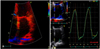
Images from the apical 4-chamber view at a high frame rate (150-180 frames per second) were obtained at end-expiratory apnea and these images were stored in a cineloop format for subsequent offline analysis. Five heartbeats were collected from each view and they were analyzed off-line with an EchoPAC Dimension system (General Electric, Horten, Norway). The peak systolic and diastolic strain rates were obtained from 2 different areas of the basal segments of the LA free wall and the inter-atrial septum on the apical 4 chamber view by the tissue Doppler strain (Fig. 1A). An appropriate velocity scale was chosen to avoid data aliasing. The narrowest image sector angle possible (usually 30° degrees) was used to achieve the maximum possible color Doppler frame rate. Careful attention was paid to keep the region of interest at the center of the ultrasound sector to ensure an alignment as close to 0° as possible to the long-axis motion. For the longitudinal measurements, a computation area of 9×2 mm with an elliptical shape was chosen. Longitudinal direction changes (measured from the apical views) for the atria are better described by the terms "rate of lengthening" in systole (the positive strain rate value) and the "rate of shortening" in diastole (the negative strain rate value). The strain rate and strain curves were calculated for all the patients over 3 cardiac cycles and then the values were averaged to obtain the mean strain rate and strain curves over 1 mean relative risk (RR) interval. End diastole was defined as the electrocardiogram (ECG) R peak, and end systole was defined as the end of the ECG T wave. The peak positive systolic and early diastolic values were calculated from the extracted curve.
Statistical analysis
All the data is expressed as means±standard deviations (SDs). The data was analyzed using standard statistical software {Statistical Package for Social Science (SPSS) package version 11.0, Chicago, IL} and comparisons of all the measurements were done using paired Student's t-test for the continuous variables and chisquare tests for the categorical variables. A p<0.05 was considered statistically significant.
Results
General characteristics of the patients
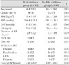
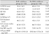
The major demographic and clinical characteristics are given in Table 1 with dividing the patients according to RAS inhibitors treatment. The total study duration was 2.1 years and none of the patients were lost to follow-up. The mean age was 66.4±8.7 years (range: 42-75 years) from group A and 66.3±8.0 years (range: 43-78 years) from group B. The global LV systolic function and, the LV chamber dimension and wall thickness were normal in all the patients with AF. The LA diameter, LA volume index, NT-pro-BNP and hs CRP showed no differences between the groups (Table 2).
Strain echocardiographic findings
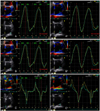
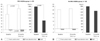
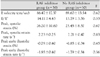
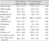
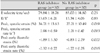
There were no significant differences of the mitral inflow parameters at baseline and at the 12-month follow-up between the groups (Table 3). There were no significant differences in the NT-pro-BNP and hsCRP levels at the baseline, but the NT-pro-BNP levels were significantly reduced more in group A (822.9±798.3 pg/mL, p<0.05) than in group B (1481.9±209.97 pg/mL) at the 12-month follow-up (Table 4). There were no significant differences of the strain parameters at baseline between the groups; however, significant increases were noted in the values of the LA strain of group A versus group B at the 12-month follow-up, specifically, there was higher mean peak systolic strain (36.71±13.63% vs. 27.21±10.49%, respectively, p=0.019), higher mean peak systolic strain rate (2.98±0.59 s-1 vs. 2.21±0.47s-1, respectively, p=0.003), higher mean peak early diastolic strain (-1.89±3.30% vs. -0.83 ±2.79%, respectively, p=0.027) and higher mean peak early diastolic strain rate (-2.32±0.77 s-1 vs. -1.77±0.25 s-1, respectively, p=0.035) in group A (Fig. 2) (Table 5). Six patients of group A (20%) and three patients of group B (10%) were converted to normal sinus rhythm during the study (Fig. 3). Because there was a larger proportion of patients with sinus conversion in group A, and the values of LA strain in normal sinus rhythm would be higher than those in AF, we compared the inter-group differences of the strain parameters with excluding the patients with normal sinus rhythm conversion. There were also significant differences between the 2 subgroups for the mean peak systolic strain (32.61±2.38% vs. 25.18±11.59%, respectively, p=0.025), the mean peak systolic strain rate (2.67±0.43 s-1 vs. 2.18±0.49 s-1, respectively, p=0.008), the mean peak early diastolic strain (-1.59±2.78% vs. -0.73±2.56%, resecively, p=0.035), and the mean peak early diastolic strain rate (-2.18±0.63 s-1 vs. -1.65±0.34 s-1, respecively, p=0.028). During follow-up, the values of the peak systolic and diastolic strain were significantly increased and the NT-pro-BNP levels were significantly decreased in group A, but not in group B (Fig. 4). Interobserver and intraobserver variability were tested with performing independent analysis by two independent observers and by repeated measurement of these segments at another occasion by the same observer. The interobserver variability was less than 20%, and the intraobserver variability was 15% for strain and 18% for the strain rate. The main reason for the interobserver variability was the different locations of the sample volume. Once the sample volume was placed on a mutually agreed location within the myocardium, the measurements became virtually identical.
Discussion
In early diastole, the atria act as conduit that passively empty during ventricular relaxation when the blood is transferred from the systemic and pulmonary veins to the ventricles.14) Thus, the atrial function during early diastole is strongly influenced by LV compliance. During ventricular systole, the atrial function as reservoirs to store blood when the AV valves are closed, and the reservoir function is influenced by atrial relaxation, ventricle contraction through the descent of the base and the atrial chamber stiffness.15) Traditionally, studies on mitral inflow (the late diastolic A wave) and pulmonary vein flow (the A reversal wave) have been used to assess LA function. TDI has recently been employed as a sensitive, reproducible tool for assessing cardiac function.16) Analyses of clinical trials suggest that TDI would be a useful non-invasive method for assessing atrial function.17)18) In a recent study, strain/rate imaging has been proposed as strong index of the atrial reservoir function, and the degree of the impairment in atrial compliance, as assessed by strain and the strain rate, seems to be strongly predictive of maintaining sinus rhythm in patients with AF.19) In our previous study, the lower values of strain/rate in the AF patients suggest decreased passive lengthening (stretching) and shortening of the atrial walls, and this is possibly because of atrial remodeling with fibrosis and the reduced compliance.13)
Electrical and structural remodeling AF is characterized by atrial dilatation and shortening of the atrial effective refractory period20)21) and this may act as perpetuators, ensuring the persistence of AF for a longer period.22) Uneven distribution of stretch on myocyte groups comes from variations in the collagen net-work and non-uniform excitation-contraction coupling.23) Electrical remodeling plays a short-lasting role in the occurrence and perpetuation of AF, and the structural atrial remodeling may be less reversible. So, the extent of structural changes is a predictor for failure of cardioversion. Acutely altered stress/strain patterns augment the synthesis of angiotensin II, which induces myocyte hypertrophy.24) Angiotensin II increases the intracellular calcium more in the atrial myocytes than in the ventricular myocytes in rats, and the density of angiotensin eceptors in the atria is generally higher than that in the ventricles. Thus, angiotensin II-induced intracellular calcium overload may play a role not only in reperfusion ventricular arrhythmias, but also in atrial electrical remodeling.25)26) Therefore, RAS inhibitors would prevent or modify atrial remodeling by means of decreasing the atrial stretch, lowering the end-diastolic left ventricular and atrial pressure, preventing atrial fibrosis and modifying the sympathetic tone, or modulating ion currents or the refractoriness. In a canine rapid atrial pacing model, pharmacologic inhibition of endogenous angiotensin II prevented early electrical remodeling (this occurs within several hours),25) with a reduction of the AF duration and structural remodeling during long-term stimulation.26) In the present study, the chronic atrial fibrillation patients who were treated with RAS inhibitors had significantly increased values of atrial peak systolic and diastolic strain/rate, as compared with the patients without treatment, and this showed that treatment with RAS inhibitors appears to preserve the LA reservoir function in AF patients without visible LA structural change. In addition, although there were too few to be statistically significant, the atrial fibrillation patients who were converted to normal sinus rhythm during the study showed more increased baseline atrial strain than did the permanent chronic atrial fibrillation patients, which would possibly indicate that higher baseline atrial strain would be a predisposing factor for conversion to normal sinus rhythm in AF patients. The results of this investigation showed agreement with a previous study that reported patients with higher atrial strain and a higher strain rate appeared to have a greater likelihood of staying in sinus rhythm.19)
The atrial biopsy specimens of AF patients have demonstrated inflammatory changes and increased fibrosis, providing evidence of the possible role of inflammation in atrial structural remodeling.21) The relation of CRP and interleukin-6 to the left atrial size and AF duration before cardioversion indicates that inflammation could have a role in the atrial structural changes, which in turn may contribute to, or be a result of atrial remodeling. An elevated BNP level has also been shown in some studies to be associated with atrial dilatation and the BNP levels have been shown by various studies to decrease after direct current cardioversion of AF and restoration of normal sinus rhythm.27) In the present study, there was no significant change in the hsCRP level, yet the NT-pro-BNP levels were significantly reduced in the patients who were treated with RAS inhibitors at the 12 month follow-up without any visible reduction of the LA size.
This study carries several limitations. Tracking of the region of interest is still suboptimal in some patients with hyperdynamic LV function and poor echocardiographic imaging. It still takes from 5 to 10 minutes to achieve satisfactory tracking, and the interobserver and intraobserver variability were relatively high because the atrial wall is too thin to be properly analyzed. Although analysis of the echocardiography parameters in AF patients would be more accurate if this was done over 5 cycles, the strain curves were calculated for all patients over 3 cardiac cycles because of the complexity. Finally, we did not consider the AF duration, so there would be differences for a shorter duration and a longer duration of AF, and the comparison between the AF patients may also have been influenced by other medications, but we tried to control the differences. To the best of our knowledge, no previous study has ever compared the change of atrial function between medical treatments by performing strain echocardiography. Our results indicate that strain echocardiography provides clinically useful information on the changes of LA function and remodeling between RAS inhibitor treatments.

 XML Download