

 PDF
PDF ePub
ePub Citation
 Print
Print


Introduction
A cardiac electrophysiologic study (EPS) is a safe procedure with a low complication rate.1) Vascular injury and deep vein thrombosis are the most common complications associated with EPS and catheter ablation.2)3) Acute myocardial infarction is a very rare complication associated with EPS.3) We report a case of unsuspected critical stenosis of the left main coronary artery treated with stenting during an EPS.
Case
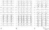
A 75-year-old man with recurrent paroxysmal chest fluttering that was accompanied by non-anginal chest discomfort was referred for an EPS. Repeated 24-hour ambulatory electrocardiograms revealed only non-sustained atrial tachycardia. A 12-lead electrocardiogram (ECG) at baseline demonstrated non-specific ST-T changes (Fig. 1A). He did not have any significant familial medical history (including diabetes mellitus, hypertension or sudden cardiac death) and he denied a history of hypertension, diabetes mellitus or hyperlipidemia. A physical examination revealed a blood pressure of 120/75 mmHg and the echocardiogram was unremarkable. The risk factors for atherosclerosis were old age and a 20 pack-year history with current smoking. The levels of total cholesterol, low density lipoprotein (LDL)-cholesterol and high density lipoprotein (HDL)-cholesterol were 169, 112 and 30 mg/dL, respectively. The other blood and urine measures were within the normal limits. An exercise stress test was not performed due to the absence of exertional angina. After an informed consent was obtained, the patient underwent an EPS in a fasting state. Three multipolar electrode catheters were introduced from the femoral vein and they were positioned in the high right atrium, the His-recording area and the right ventricle. The basic intervals were a sinus cycle length of 1,090 ms, an AH interval of 60 ms and an HV interval of 45 ms. There was no evidence of an antegrade or retrograde bypass tract. There was only retrograde nodal conduction without any antegrade dual atrioventricular (AV) node physiology. Supraventricular tachycardia was not induced at baseline. Sustained atrial fibrillation with a duration of more than 10 minutes was reproducibly induced by double atrial extrastimulation via an infusion of isoproterenol at 1 µg/min. He complained of severe persistent chest pain during the atrial fibrillation. A 12-lead ECG demonstrated widespread ST depression in the precordial and inferior leads and ST elevation in aVR and V1 (Fig. 1B). The ST seg ment deviations were more exaggerated after normal sinus rhythm was restored by electrical cardioversion (Fig. 1C). We performed emergency coronary angiography and intervention via the right radial artery after informed consent was obtained. Left coronary angiography showed 99% stenosis in the distal left main coronary artery (LMCA) with thrombolysis in myocardial infarction (TIMI) trial grade 2 antegrade flow, and there was 50% stenosis at the mid-left anterior descending artery (LAD) (Fig. 2A). Right coronary angiography showed 70% stenosis at the posterolateral branch. Intravenous infusion of heparin in conjunction with a loading dose of clopidogrel and aspirin was given. Coronary intervention was then performed with placement of a 3.0×23 mm sirolimus-eluting stent (CYPHER™; Cordis Corporation, Miami Lakes, FL, USA) in the LMCA after performing balloon predilatation, and TIMI-3 flow was then established and maintained. A 3.5 mm, high-pressure, non-compliant balloon was used for post-stent dilation (Fig. 2B). It took 40 min from the detection of the ECG change to restoration of TIMI-3 flow in the LMCA. The hemodynamic status was stable during the entire coronary intervention. After the procedure, the level of creatine kinase-MB (CK-MB) rose to 74.6 IU/L. Echocardiography after the procedure showed preserved left ventricular (LV) systolic function with an akinetic LV apex and anteroseptum. He recovered without complication and was discharged to home on aspirin, clopidogrel, warfarin, diltiazem and artovastatin. He has remained free of paroxysmal palpitations and angina with these medications for one year. Follow-up coronary angiography after one year showed a patent stent in the LMCA and no interval change of the remaining arteries (Fig. 2C).
Discussion
A cardiac EPS is an established diagnostic procedure with proven safety.1) According to a large-scale study, the most common vascular complications are vascular injury and deep vein thrombosis.2)3) Acute myocardial infarction is a very rare complication associated with EPS.3)
Early identification of stenosis of the LMCA is crucial to plan a therapeutic strategy for patients with acute chest pain. Elevation of the ST segment in the aVR lead is associated with multivessel disease or LMCA disease in patients with acute coronary syndrome,4)5) and in patients with acute occlusion of the LMCA, especially when the ST segment elevation in the aVR lead is greater compared to that of lead V1.6)7) The mechanism of ST segment deviation in the same direction as acute ischemia and occlusion of LMCA is unknown.
Percutaneous revascularization of the LMCA has shown excellent clinical results and low morbidity in select patients. 8)9) Even though the standard treatment of LMCA stenosis is coronary artery bypass graft surgery,10) percutaneous revascularization of the LMCA may be a lifesaving procedure in the setting of ongoing ischemia as a bail-out procedure for treating iatrogenic LMCA injury in the catheterization laboratory.11-13)
Myocardial ischemic symptoms in the elderly are often absent or atypical compared with young patients.15) Asymptomatic myocardial ischemia can be documented during ambulatory ECG monitoring, but this was not found in the present case.14) Even though the patient did not complain of exertional angina, a stress myocardial imaging test might have revealed clinically unsuspected significant coronary artery disease. Considering the atypical symptoms of myocardial ischemia and ischemic complications in the elderly, performing a stress myocardial imaging test should be seriously considered before conducting an EPS in elderly patients.3)15)
In conclusion, we report here a case of unsuspected critical LMCA stenosis that was treated with stenting during an EPS. To prevent this potentially catastrophic complication, stress myocardial perfusion imaging should be strongly considered in those elderly patients who do not have ischemic symptoms before an EPS.

 XML Download