

 PDF
PDF ePub
ePub Citation
 Print
Print


Abstract
Background and Objectives
Certain types of arrhythmias have gender differences. Women have a higher incidence of drug-induced QT prolongation than in men. However, there are no reports regarding gender-related differences of the P wave signal averaged electrocardiogram (PWSAE), based on the risk of atrial fibrillation (AF). PWSAE has been recognized as a diagnostic tool for identifying the risk of paroxysmal atrial fibrillation (PAF). We therefore investigated the influence of gender in the parameters of PWSAE and in the risk of AF.
Subjects and Methods
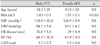
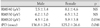
We recorded 100 PWSAEs in apparently healthy Korean subjects (53 men and 47 women), aged 20 to 79 years.
Results
The mean age of the male subjects was 38.2 years and the mean age of the female subjects was 43.2 years (p=0.19). The body surface area (BSA) were larger in men (1.83 m2 vs. 1.53 m2, p<0.05). In men, the filtered P wave duration (fPD) was longer than in women (136.8 msec vs. 125.2 msec, p<0.05). The root mean square voltage in the terminal 20 ms of the filtered P wave (RMS20) was 5.9 µV in women and 4.5 µV in men (p<0.05).
Conclusion
Men have a longer fPD and lower RMS20 than women. The BSA showed a positive correlation with fPD and a negative correlation with RMS20. This study suggests that BSA is an important factor for fPD and RMS20. In addition, as men have a larger BSA as compared with women, we suspect that men have a higher risk of AF as compared with women.
Figures and Tables
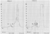
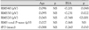
Fig. 2
Correlation between age, body surface area and the filtered P-wave duration (fPD). A: there was a weak positive correlation between fPD and body surface area (r=0.260, p=0.010). B: no correlation was observed between RMS20 and age. RMS20: root mean square of the terminal 20 milliseconds.
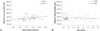
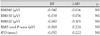
Fig. 3
Correlation between age, body surface area and RMS20. A: there was a negative correlation between RMS20 and the body surface area (r=-0.348, p<0.001). B: no correlation was observed between RMS20 and age. RMS20: root mean square of terminal 20 milliseconds.
References
1. Kannel WB, Abbott RD, Savage DD, McNamara PM. Epidemiologic features of chronic atrial fibrillation. N Engl J Med. 1982. 306:1018–1022.
2. Josephson ME, Zimetbaum P. Kasper DL, Braunwald E, Fauci AS, editors. The Tachy arrhythmias. Harrison's Principles of Internal Medicine. 2005. 16th ed. McGraw-Hill;1345–1347.
3. Tanikawa M, Fukatani M, Kenoe A, Isomoto S, Kadena M, Hashiba K. Prolonged and fractionated right atrial electrograms during sinus rhythm in patients with paroxysmal atrial fibrillation and sick sinus node syndrome. J Am Coll Cardiol. 1991. 17:403–408.
4. Centurion OA, Isomoto S, Fukatani M, et al. Relationship between atrial conduction defects and fractionated atrial endocardial electrograms in patient with sick sinus syndrome. Pacing Clin Electrophysiol. 1993. 16:2022–2033.
5. Kim JK, Kim JS, Lee HH, et al. Analisis of P wave signal-averaged electrocardiogram in patients with paroxysmal atrial fibrillation. Korean Circ J. 2002. 32:146–154.
6. Hwang GS, Kim YH, Lee HS, et al. Electrical remodeling in human atrial fibrillation influence post-cardioversion atrial mechanical dysfunction and early relapse. Korean J Cardiovasc Dis. 2000. 1:90–96.
7. Ehlert FA, Zaman N, Steinberg JS. Immediate and short-term reproducibility of the P wave signal-averaged electrocardiogram. Pacing Clin Electrophysiol. 1997. 20:1636–1645.
8. Stafford PJ, Cooper J, Fothergill J, Schlindwein F, de Bono DP, Garratt CJ. Reproducibility of the signal-averaged P wave: time and frequency domain analysis. Heart. 1997. 77:412–416.
9. Dilaveris PE, Gialafos JE. P-wave duration and dispersion analysis: methodological considerations. Circulation. 2001. 103:E111-1.
10. Danford DA, Stelling JA, Kugler JD, et al. Signal-averaged electrocardiography of the terminal QRS in healthy young adults. Pacing Clin Electrophysiol. 1989. 12:1712–1716.
11. Ohe T, Matsuhisa M, Kamakura S, et al. Relation between the widening of the fragmented atrial activity zone and atrial fibrillation. Am J Cardiol. 1983. 52:1219–1222.
12. Simpson RJ Jr, Foster JR, Gettes LS. Atrial excitability and conduction in patient with interatrial conduction defects. Am J Cardiol. 1982. 50:1331–1337.
13. Dilaveris PE, Gialados EJ, Sideris SK, et al. Simple electrocardiographic marker for the prediction of paroxysmal idiopathic atrial fibrillation. Am Heart J. 1998. 135:733–738.
14. Andrikopoulos GK, dilaveris PE, Richter DJ, Gialados EJ, Synetos AG, Gialafod JE. Increased variance of P wave duration on the electrocardiogram distinguishes patient with idiopathic paroxysmal atrial fibrillation. Pacing Clin Electrophysiol. 2000. 23:1127–1132.
15. Guidera S, Steinberg JS. The signal-averaged P wave duration: a rapid and noninvasive marker of risk of atrial fibrillation. J Am Coll Cardiol. 1993. 21:1645–1651.
16. Stafford PJ, Turner I, Vincent R. Quantitive analysis of signal-averaged P waves in idiopathic paroxysmal atrial fibrillation. Am J Cardiol. 1991. 68:751–755.
17. Kumagai K, Akimitsu S, Kawahira K, et al. Electrophysiological properties in chronic lone atrial fibrillation. Circulation. 1991. 84:1662–1668.
18. Steinberg JS, Zelenkofske S, Wong SC, Gekernt M, Sciacca R, Menchavez E. The value of the P-wave signal averaged ECG for predicing atrial fibrillation after cardiac surgery. Circulation. 1993. 88:2618–2622.
19. Kim W, Shin DG, Hong GR, et al. Signal averaged P wave dispersion: a new marker for predicting the risk of paroxysmal atrial fibrillation. Korean Circ J. 2002. 32:339–348.
20. Ehlert FA, Korenstein D, Steinberg JS. Evaluation of P wave signal-averaged electrocardiographic filtering and analysis methods. Am Heart J. 1997. 134:985–993.
21. Yamada T, Fukunami M, Ohmori M, et al. Characteristics of frequency content of atrial signal-averaged electrocardiograms during sinus rhythm in patients with paroxysmal atrial fibrillation. J Am Coll Cardiol. 1992. 19:559–563.
22. Babaev AA, Vloka ME, Sadurski R, Steinberg JS. Influence of age on atrial activation as measured by the P-wave signal-averaged electrocardiogram. Am J Cardiol. 2000. 86:692–695.
23. Faggiano P, D'Aloia A, Zanelli E, Gualeni A, Musatti P, Giordano A. Contribution of left atrial pressure and dimension to signal-averaged P-wave duration in patients with chronic congestive heart failure. Am J Cardiol. 1997. 79:219–222.
24. Cheema AN, Ahmed MW, Kadish AH, Goldberger JJ. Effects of autonomic stimulation and blockade on signal-averaged P wave duration. J Am Coll Cardiol. 1995. 26:497–502.
25. Ehlert FA, Steinberg JS. The P wave signal-averaged ECG. J Electrocardiol. 1995. 28:Suppl. 33–38.
26. Fukunami M, Yamada T, Ohmori M, et al. Detection of patient at risk for paroxysmal atrial fibrillation during sinus rhythm by P wave-triggered signal-averaged electrocardiogram. Circulation. 1991. 83:162–169.
 XML Download