

 PDF
PDF ePub
ePub Citation
 Print
Print


Introduction
It is well known that atherosclerosis has a complex pathogenesis that involves local factors in the vessel wall such as vascular inflammation and also systemic factors such as dyslipidemia and insulin resistance.1) Peroxisome proliferator-activated receptor (PPAR) agonists have been regarded as one of the promising antiatherogenic agents to control both local and systemic atherogenic factors.2) Experimental animal and in vitro studies and some clinical studies too have demonstrated that PPAR agonists limit vascular inflammation and improve insulin resistance and the lipid profiles.3-5) Fibric acid derivatives are known as PPAR-α agonists, and they are already being clinically used for lowering lipid levels. Previous clinical trials of fibrates have demonstrated their beneficial effects on cardiovascular clinical outcomes, and particularly among patients suffering with metabolic syndrome and diabetes.6-10) However, the recent large randomized clinical studies11)12) that have evaluated the more potent PPAR-α agonist have questioned the beneficial effects of PPAR-α agonists, and these studies have presented a variety of debatable issues to clinicians who have prescribed PPAR-α agonists for such diseases.
This review 1) describes the PPAR biology and the mechanism of action of PPAR-α agonists and 2) it summarizes the results of clinical trials of PPAR agonists on atherosclerotic diseases, with a special focus on their safety and efficacy.
Brief Peroxisome Proliferator-Activated Receptor Biology
Issemann and Green13) discovered that clofibrate activated an orphan nuclear receptor, which they named the PPAR.
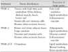
This name was based on the observation that these agents induce the proliferation of peroxisomes, a cell organelle, in rodents. The PPAR family consists of 3 members, namely, PPAR-α, PPAR-γ and PPAR-β/δ, which all share approximately 60-80% homology in their ligand-binding domains (LBD) and DNA-binding domains (DBD). Each subtype of PPAR has a distinct tissue distribution, target gene, individual encoding gene and physiological action (Table 1).
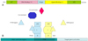
PPARs have 5 domains14) (Fig. 1A): 1) the LBD, to which the specific PPAR agonist binds; 2) the N-terminal AF2 domain, which can be activated by phosphorylation through the mitogen-activated protein (MAP) kinase sites via ligand-independent activation; 3) the DBD, which interacts with specific PPAR response elements (PPRE) in the promoter region of the PPAR-activated target genes; 4) the C-terminal AF2 domain, which, in response to ligand binding, undergoes a permissive conformational change that's required for transcriptional activation; 5) the hinge domain, whose function is not well understood, but whose structure is very flexible and it may be crucial for efficient binding of the DBD.
PPAR activation via the binding of LBD to specific ligands leads to its heterodimerization with the retinoid X receptor (RXR), and it undergoes a conformational change in the AF2 domain, which facilitates the release of co-repressors and the recruitment of coactivators(Fig. 1B).15) In contrast to the positive transcriptional regulation of PPAR as described above, PPAR activation can also repress the transcription of the target genes by unknown mechanisms.16) This repressive action of PPARs is the major mechanism that underlies its anti-inflammatory action.
As described above, transcriptional regulation by PPAR agonists requires multiple levels of control as follows: PPAR ligand with synthetic ligands and natural ligands, corepressors and coactivators, homology of the LBD between each subtype of PPAR, and many known and unknown target genes in multiple organs. In fact, synthetic agonists can activate only a specific subtype of PPAR. For example, fibrates bind PPAR-α, which controls lipid metabolism, and thiazolinediones bind PPAR-γ, which controls hyperglycemia. However the effects of synthetic PPAR agonists in vivo differ from those of PPAR itself because the synthetic agonists may be greatly influenced by natural ligands in vivo, and these natural ligands are as yet unknown.5) Corepressors and coactivators form a large, diverse family with members such as nuclear corespressor,17) PPAR-binding protein,18) PPAR-γ coactivator19) and cAMP response element-binding protein.20) PPAR activation by synthetic ligands may modulate a large number of genes, and some of which produce unknown effects. Particularly, the dual or broad PPAR agonists may be more dangerous.21) PPAR activation may be beneficial in one organ, but harmful in another organ. The clinical application of PPAR agonists for therapeutic targets may require a more comprehensive understanding about PPAR biology and this must be carefully approached.
Effects of Peroxisome Proliferator-Activated Receptor-α on Lipid Metabolism, the Vessel Wall and the Heart
PPAR-α is metabolically active in the liver, heart, kidney, skeletal muscle and brown fat.22)23) It is also present in all vascular cells, including endothelial cells, smooth muscle cells and monocytes/macrophages.24-26) The effects of PPAR-α include hypolipidemic action, an anti-inflammatory effect on the vascular wall and metabolic effects on the myocardium.
Hypolipidemic action
PPAR-α agonists, i.e., the fibrates (clofibrate, gemfibrozil, fenofibrate, benzafibrate, and ciprofibrate), have been used as lipid-lowering agents for over 40 years, and this is mainly due to their action of lowering triglyceride (TG) levels and raising high density lipoprotein cholesterol (HDL-C) levels.
The lowering of TG levels in plasma after PPAR-α activation is attributed to the following mechanisms: 1) The increased diversion of fatty acids into β-oxidation, thereby limiting their availability for TG and very low-density lipoprotein (VLDL) synthesis,27) 2) the inhibition of apo CIII, which is an inhibitor of lipoprotein lipase (LPL)28) and this increases LPL activity,29) which enhances the hydrolysis of TG-rich particles and improves the uptake of their remnants.
The increased HDL-cholesterol levels in plasma after PPAR-α activation may be explained by the following factors: 1) increased production of apo AI and apo AII, which are the major HDL components,30)31) 2) increased transfer of the other surface components of triglyceriderich particles to HDL by enhancing the LPL activity,32) and 3) an enhanced APT-binding cassette transporter A1 (ABCA1) expression as a result of PPAR-α activation.33)
Effects on the vascular wall
The anti-inflammatory action of PPAR-α on all vascular cells has been studied and reported on, and most notably by both in vitro and in vivo studies.4) PPAR-α activators inhibit the production of inflammatory response markers such as endothelin-1, vascular adhesion molecule-1 (VCAM-1), interleukin (IL)-6 and tissue factors in endothelial cells, smooth muscle cells and macrophages.36-45) In patients with dyslipidemia, PPAR-α agonists reduce the levels of inflammatory markers such as IL-6, fibronogen, C-reactive protein, serum amyloid A, plasminogen, α2-macroglobin, interferon-γ, IL-2, tumor necrosis factor-α and IL-1β.42)46-51) These effects of PPAR-α agonists on the vessel wall may explain their cardiovascular protective effects that extend beyond their lipid lowering effect.10)
Effects on myocardium
The cardiac metabolic effects of PPAR-α activation are less well defined. Although the fetal heart obtains most of its energy from glucose and lactate, the adult heart obtains its energy from PPAR-α-dependent fatty acid oxidation, as well as from glucose and lactate, in order to meet its energy demands under varying dietary and physiological conditions.27)52) In a murine model of pressure induced cardiac hypertrophy, PPAR-α was observed to be down-regulated,52)53) which indicates that the cardiac metabolism shifted from fatty acid oxidation to glucose utilization. PPAR-α activation in this model resulted in severe left ventricular dysfunction. It is suggested that PPAR-α downregulation in a hypertrophic heart may be an adaptive process that is essential for maintaining normal heart function.54) The significance of PPAR-α activation on human cardiac hypertrophy has not yet been established.
In the hearts of patients with uncontrolled diabetes, impaired glucose utilization results in almost exclusive use of fatty acid oxidation to provide for the ATP needs of the myocardium.55) In that case, PPAR-α activation may theoretically include conflicting effects on the myocardial energy metabolism. The beneficial effect is that PPAR-α agonists cause a hypolipemic state that may reduce the amount of fatty acids, which is a substrate of fatty acid oxidation, delivered to the myocardium. However, another potentially harmful effect is that PPAR-α activation triggers the shift from glucose utilization to fatty acid oxidation as an energy source. Increased fatty acid oxidation may lead to an increased oxygen demand, and a high uptake of fatty acid by the myocardium may lead to lipid accumulation in the myocardium, which predisposes it to systolic dysfunction and heart failure. Therefore, questions have been raised regarding the net effect of PPAR-α activation on the diabetic myocardium. In a diabetic murine model, PPAR-α activation revealed that the reduction of the delivered substrate was more important than the energy switch.56) The treatment with PPAR-α agonist normalized the free fatty acid, TG and glucose levels; moreover, it also reduced myocardial fatty acid oxidation by 50% and increased glucose utilization. In contrast, the cardiac-specific overexpression of PPAR-α in non-diabetic mice revealed increased fatty acid oxidation and decreased glucose utilization in the myocardium, and this induced a diabetic-type cardiomyopathy in otherwise normal mice. In diabetic patients, the effects of PPAR-α activation on myocardial metabolism are still under debate and this requires further study.
In the ischemic heart, glucose utilization requires less oxygen than fatty acid oxidation; further, it does not worsen acidosis in the ischemic myocardium. Therefore, inhibitors of fatty acid oxidation such as trimetazidine are being used in clinical practice.57) Theoretically, PPAR-α agonist may compromise this anti-anginal effect of fatty acid oxidation inhibitors. However, since severe ischemia itself can turn off fatty acid oxidation, the role of PPAR-α in the ischemic human myocardium needs to be clearly defined. The pretreatment of an infarction and an ischemia-reperfusion model with fibrates demonstrated a reduction in the size of infarction and improved postischemic contractile function because PPAR-α activation had an anti-inflammatory effect.58)59) These results suggest that pre-ischemic treatment with PPAR-α agonist may limit the ischemic damage; however, post-ischemic treatment might be theoretically harmful if it shifts the cardiac metabolism from glucose utilization to fatty acid oxidation.
Summary of the Important Clinical Trials That Used Peroxisome Proliferator-Activated Receptor-α Agonists
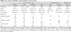
Several large clinical studies have investigated the potential cardioprotective effects of PPAR-α agonists, and particularly the fibrate derivatives. These studies have exhibited a wide range of results (Table 2):
No beneficial results or harmful results: the World Health Organization (WHO) cooperative trial, the Coronary Drug Project (CDP) trial, the Lower Extremity Arterial Disease Event Reduction Study (LEADER), and the Bezafibrate Infarction Prevention Trial (BIP)
Beneficial results: the Helsinki Heart Study (HHS) and the Veteran's Affairs-HDL Intervention Trial (VA-HIT)
Mixed results: the Fenofibrate Intervention and Event Lowering in Diabetes (FIELD) study
World health organization cooperative trial
Male patients (n=15,745) without coronary heart disease (CHD) were enrolled and followed up for a mean period of 5.3 years.60) Of the major CHD events, only the incidence of non-fatal myocardial infarction (MI) was significantly reduced in the fibrate group (relative risk reduction: 20%). There was no difference in the number of deaths due to cardiac causes between the groups. The overall mortality was higher in the clofibrate group; this was attributed to diseases of the liver, intestines and gall bladder.61)
Coronary drug project trial
Male patients (n=8, 341) with one or more MI attacks were randomized to 1 of 6 treatment groups and they were followed up for 5-8.5 years.62) Follow up was discontinued early in 3 of those 6 groups due to the increased incidence of cardiac events. The remaining 3 groups were comprised of patients who were treated with clofibrate, niacin and placebo. Overall, there was no statistically significant difference in the total mortality, and the incidence of nonfatal MI and cardiac deaths. Furthermore, this trial showed a statistically nonsignificant increase in thromboembolism, angina, intermittent claudication, cardiac arrhythmia and gall stones, along with an increase of the nonfatal cardiovascular events.
Lower extremity arterial disease event reduction trial
The treatment of 1,568 men with either bezafibrate or placebo demonstrated that the incidence of CHD and stroke was not reduced in the bezafibrate treated group.63) The beneficial effects on non-fatal cardiac events were the greatest in men aged <65 years at study entry; a beneficial effect on all coronary events was also observed in this group of patients. There were no significant effects in men aged ≥65 years. There was no difference between the 2 groups with respect to all causes of deaths. Bezafibrate only reduced the severity of intermittent claudication for up to 3 years.
Bezafibrate infarction prevention trial
In this trial,7) 3,090 patients with CHD and dyslipidemia (total cholesterol: 180-250 mg/dL, HDL-C≤45 mg/dL, TG≤300 mg/dL and LDL-C≤180 mg/dL) were randomized to receive either bezafibrate or a placebo. After follow up (mean duration: 6.2 years), there was no difference in the incidence of fatal and nonfatal MI or sudden death between the 2 groups. The total mortality and noncardiac mortality were also similar, and the incidences of adverse events and cancer were equally distributed. Only the subgroup with a high baseline TG level (≥200 mg/dL) enjoyed the reduction in the cumulative probability of myocardial infarction or sudden death by bezafibrate administration.
Helsinki heart study
This primary prevention trial6) with using gemfibrozil was performed for 5 years on 4,081 asymptomatic middle-aged men (age range: 40-55 years) with primary dyslipidemia (non-HDL cholesterol≤200 mg/dL). This trial demonstrated a 34% reduction in the incidence of overall cardiac events, including fatal and nonfatal MI and cardiac death. This effect is more marked in patients suffering with diabetic and metabolic syndrome.64) No difference was noted in the mortality due to all-causes; however, an increase was noted in the deaths due to noncoronary causes.
A substudy of the HHS,65) which employed the males excluded from the primary prevention cohort due to a history of MI, angina or prior ECG changes, also showed no difference between the gemfirozil group and the placebo group
Veteran's administration-HDL intervention trial
In this study,8) 2,531 men with CHD, low HDL (≤40 mg/dL), and moderately elevated LDL-C (≤140 mg/dL) were randomized and treated with either gemfibrozil or placebo for 5.1 years. Although the LDL-C levels did not significantly differ between the groups, the gemfibrozil group showed a significant reduction in the risk of nonfatal MI and cardiac death (relative risk reduction: 22%). Further, a 24% reduction was noted in the combined outcome of death from CHD, nonfatal MI and stroke. There were no significant differences in the rates of coronary revascularization, hospitalization for unstable angina, death from any cause and cancer. The experts concluded that the beneficial effect of gemfibrozil on cardiovascular events may have been driven largely by the characteristics of the enrolled group in the VA-HIT as the subjects were mostly patients with diabetes and/or metabolic syndrome, along with the lipid inclusion criteria.9)10)
Fenofibrate intervention and event lowering in diabetes
This study11) was comprised of a mixed population of 9,795 low-risk "primary prevention" and medium-risk "secondary prevention" diabetic patients. Fenofibrate treatment showed a significant 24% reduction in the incidence of non-fatal MI and a non-significant increase in CHD mortality. The total cardiovascular disease events were significantly reduced, and this was mainly due to a reduction in coronary revascularization and nonfatal MI. The total mortality was similar in both the groups. No benefit was observed in the "secondary prevention" subgroup. Unexpectedly, fenofibrate treatment was associated with a lower incidence of microvascular complications such as albuminuria progression and retinopathy that required laser treatment. However, except for a slight increase in the incidence of pancreatitis and pulmonary embolism, no other significant adverse effects were noted. The authors concluded that the higher use of statins in the placebo group might have masked a moderately larger treatment benefit.
More potent and selective peroxisome proliferator-activated receptor-α agonist, LY518674, trial
The novel selective PPAR-α agonist, LY518674, is approximately 10,000 times more potent than fenofibrate. This study12) aimed at examining the safety and efficacy of LY518674 among 2 populations of patients with atherogenic dyslipidemia and hypercholesterolemia. In a trial of 309 patients with atherogenic dyslipidemia, LY518674 and fenofibrate both individually outperformed the placebo, with no significant differences being noted between the 2 PPAR-α agonists. LY518674 also raised the LDL level in a dose-dependent fashion and it did so much more than fenofibrate. In the other trial that was comprised 304 hypercholesterolemic and statin-naive patients, LY518674 and atorvastatin each significantly reduced the TG and LDL levels, and they both increased the HDL levels. For the atorvastatin recipients, the addition of LY518674 further increased the HDL levels (by 1-12%) and reduced the TG levels; however, it had little effect on the LDL levels. LY518674 and fenofibrate demonstrated evidence of increasing the serum creatinine levels; this effect was substantial in some cases.
The novel PPAR-α agonist was not better than fenofibrate or statin monotherapy in achieving the intended improvement of the lipid profile, and it appeared to worsen renal function.
Efficacy from the Clinical Trials of Peroxisome Proliferator-Activated Receptor-α Agonist
In the LOCAT66) (Lopid Coronary Angiography Trial), the DAIS67) (Diabetes Atherosclerosis Intervention Study) and other clinical studies,68) fibrates have demonstrated a reduction in the rates of progression of coronary atheromas as measured by quantitative angiography. However, the cardioprotective effects of PPAR-α agonists were not consistent in the individual clinical trials.
When comparing the results of 2 initial large-scale trials of clofibrate, i.e., the CDP trial62) and the WHO cooperative trial,60) the benefit for reducing CHD events was not significant in the former, but it was significant in the latter (relative risk reduction: 20%, p<0.05). However, both studies showed a significant increase in the total mortality (p<0.05). In the BIP trial,7) bezafibrate treatment showed an 11% relative reduction in the risk of CHD events, but this was not statistically significant (p=0.26). However, post hoc analysis revealed a significant reduction in the risk of CHD events (relative risk reduction: 22%, p=0.02) in those patients with higher baseline TG levels (≥200 mg/dL). The LEADER trial63) showed no beneficial effect of bezafibrate treatment on cardiovascular disease except for a reduction in the risk of nonfatal MI in young men. All the patients of the LEADER trial were also treated with statins, and the outcome results were compounded by the effects of statins. In contrast to the disappointing results of the clofibrate and bezafibrate trials, the gemfibrozil trials (the HHS and VA-HIT) demonstrated a statistically significant reduction in the incidence of cardiovascular events. The VA-HIT results helped to stimulate considerable anticipation for the results of the FIELD study11) that used fenofibrate for treating first or recurrent cardiovascular events in patients with type 2 diabetes. However, the results of the FIELD study showed a statistically nonsignificant difference between the treatment and placebo groups with respect to reducing the primary endpoints, including CHD death and non-fatal MI, and there was a slightly increase of the total and cardiovascular mortality. In the "primary prevention" group, fenofibrate treatment resulted in a cardioprotective effect only against the secondary endpoints such as nonfatal MI and revasularization procedures. This major discrepancy between the results of the VA-HIT and FIELD has led to several debates and it has raised many questions.
First, the cardioprotective effects of gemfibrozil in the HHS and VA-HIT were greater than those of other fibrates in the WHO cooperative trail, the BIP trial and the FIELD study. Some researchers suggest that gemfibrozil has less severe adverse effects than do the other fibrates. Gemfibrozil does not increase the levels of homocysteine and creatinine to the same extent that has been observed with using the other fibrates.69)70) Indeed, in the VA-HIT study, the benefit of gemfibrozil due to the increase in HDL-C could account for only 20% of the CHD reduction in terms of the lipid changes.71) This result suggests that gemfibrozil itself has other beneficial effects that extend beyond its lipid-modulating effect.
Second, the cohort of the FIELD study had different characteristics from those subjects of the VA-HIT. The baseline HDL-C levels of the FIELD cohort were relatively high (mean HDL-C: 42 mg/dL); this might have masked the cardioprotective effects of fenofibrate. The VA-HIT was designed to evaluate the effect of gemfibrozil in patients who were not using statin. However, the control group of the FIELD study had an asymmetrical higher statin drop-in due to the LDL-C-lowering effect of fenofibrate, which might have also masked the beneficial effects of fenofibrate. In contrast, some might argue that the beneficial effect in the FIELD study might be attributed to the modest LDL-C low-ering effect of fenofibrate.72)
Third, some suggest that the potency of PPAR-α agonist might have had an influence on the cardiac outcome results. PPAR is an extremely critical transcriptional regulator, and a number of factors can affect it. In view of the mechanism of action of PPAR agonists, it may be very difficult to get clinically beneficial effects by using only a synthetic agent, which may result in beneficial and/or unwanted effects. Theoretically, the final net effect of both the beneficial and unwanted effects must be considered to be more important. Gemfibrozil is less potent than fenofibrate. The result of the VA-HIT suggests that more potent PPAR activation may not be essential for achieving clinical benefits. Furthermore, LY518674, which is a potent PPAR-α agonist, showed no better results in achieving an improved lipid profile than did fenofibrate or statin monotherapy.12)
Fourth, we do not have a clear answer as to whether combination therapy with statin and fibrate may lead to a beneficial cardiac outcome. Combination therapy may achieve an optimal improvement in the lipid profile and offer greater cardiovascular risk reduction; however, it might increase the risk of rhabdomyolysis.73) When the Action to Control Cardiometabolic Risk in Diabetes (ACCORD) study is completed, good evidence regarding combination therapy will become available. In the ACCORD study design, fenofibrate is added to baseline 20-40 mg simvastatin administration in 5,800 patients in a 2×2 design trial with ultratight glycemic control versus the usual glycemic control.
Safety Issues from the Clinical Trials of Peroxisome Proliferator-Activated Receptor-α Agonist
The safety issues of PPAR agonists have recently entered the spotlight since Nissen et al.74) argued about the potentially cardiotoxic effects of rosiglitazone, a PPAR-γ agonist that's widely used as a hypoglycemic agent. Fortunately, none of the failed PPAR agonists that showed severe side effects were pure PPAR-α or PPAR-α-preferential dual agonists when they tested on the human PPAR isoforms.75) Indeed, for most of the failed PPAR agonists, their apparent affinity for PPAR-γ is higher than their affinity for PPAR-α. Furthermore, because the PPAR-α agonists, i.e., fibrates, have been used for 40 years, they are generally considered as safe drugs with only a few side effects. However, the safety issues of PPAR-α agonists include increased total mortality, muscle toxicity such as myopathy or rhabdomyopathy, increased levels of plasma creatinine and homocysteine, and lithogenicity. We will briefly discuss here the unwanted effects of PPAR-α agonists based on the results of clinical trials.
Increased total mortality
Although clinical trials have demonstrated that treatment with fibrates reduces nonfatal MI, all the clinical trials of fibrates, other than the VA-HIT, showed either no increase or a slight increase in the total mortality due to noncardiovascular causes in the treatment group. More recently, the FIELD study11) showed a nonsignificant increase in the cardiovascular, noncardiovascular, coronary and total mortality associated with fenofibrate treatment. However, the relationship between total mortality and fibrate treatment was not statistically powerful, and the increased mortality was not attributable to any specific causes of death (such as invasive cancer).76) It is not known whether the increased or unchanged rates of mortality are a chance finding or if they are side effects of this drug. However, concern could be raised that the unwanted effects of PPAR-α agonists might be related to the increased mortality and this should be resolved in the future.
Rhadomyolysis, myopathy and myalgia
Monotherpy as well as combined therapy with fibrates may be associated with cases of myopathy, myalgia, and in extremely rare cases, with rhabdomyolysis.77) The mechanism of fibrate-induced muscle toxicity is unknown; however, it may be explained on the basis of PPAR-α-dependent and compound-dependent actions. Severe myopathy in mice correlates with an increased expression of the LPL-target protein of PPAR-α in skeletal muscles.78) Some authors suggest that the energy imbalance due to increased fatty acid oxidation and decreased TG in skeletal muscle may cause the degradation of muscle protein.79)80) Muscle toxicity has been noted with both gemfibrozil and fenofibrate administration, but it is more frequent with the former (rhadomyolysis incidence: 59.6/million and 5.5/million, respectively).81)
In clinical practice, combination therapy with both fibrate and statin definitely increases the risk of rhadomyolysis and myopathy. Furthermore, combined therapy with gemfibrozil plus statin is considered more risky than that with fenofibrate and statin. Recent reviews of the Food and Drug Administration's Adverse Event Reporting System database reported that the rate of myopathy with combined gemfibrozil and statin therapy was 33 times more than that with combined fenofibrate and statin therapy.73)81) This difference is attributed to the ability of gemfibrozil to increase the plasma statin level to some extent because it uses enzymes from the same family of glucuronidation enzymes as do the statins; however, fenofibrate uses enzymes from a different enzyme family.73)
Increased plasma creatinine
A modest and reversible elevation of the plasma creatinine level has been reported in clinical trials that have used bezafibrate82)83) and fenofibrate82)84)85) and less commonly, in trials that have used gemfibrozil.84) Some authors have suggested that creatininemia may be related to a PPAR-α activation-induced inhibition of the cyclooxygenase (COX-2) gene expression in the kidney86) and the resultant decrease in the synthesis of the vasodilator prostaglandin.87) It's interesting that the use of tesaglitazar, a dual PPAR-α/γ agonist, was discontinued because of severe renal toxicity.88) However, recent reports suggest that fibrate-induced creatininemia was not associated with the glomerular filtration rate (GFR), which is assessed by using the insulin or creatinine clearance,89) and this was attributed to increased creatinine production and particularly from muscle tissue. Although the harmful renal effect of PPAR-α agonist is uncertain, using these drugs clinically in patients with renal insufficiency should be carefully considered.
Increased plasma homocysteine
Fenofibrate, ciprofibrate and bezafibrate all increase the plasma homocysteine concentration of dyslipidemic and diabetic patients,90-93) whereas gemfibrozil does so to a lesser extent. Although the exact mechanism for this is not known, fibrate treatment did not cause homocysteinemia in PPAR-α knockout mice; however, it led to homocysteinemia in wild mice.94) This finding suggests that fibrate-induced homocysteinemia may be related to PPAR-α activation. The clinical significance of hyperhomocysteinemia remains a matter of discussion. The DAIS67) showed that fenofibrate-induced homocysteinemia did not attenuate its beneficial effect for inhibiting the progression of atherosclerosis.
Increased gallstones
There was a marked association between clofibrate and gallstones in the WHO cooperative trial60)61) and the CDP trial,62) and the latter trial showed a 2-3 times higher rate of cholecystectomy in the treatment group as compared with the placebo group. A possible mechanism of lithogenicity could be that PPAR-α agonists reduce the expression of cholesterol 7α-hydroxylase and sterol 27-hydroxylase, and this results in an increased cholesterol saturation index.95)
It is unknown whether gemfibrozil or fenofibrate can be a causative factor for gallstones because the rates of cholecystectomy in the clinical trials, including the HHS, VA-HIT and FIELD study, were not reported.
Conclusions
Considering the benefits and safety concerns, the PPAR-α agonists (fibrates) that are currently in use may definitely be beneficial for decreasing the rate of clinical cardiovascular events in a specific population, and particularly among those patients suffering with metabolic syndrome and atherogenic dyslipidemia. Large-scale long-term clinical trials of PPAR-α agonists have shown that these drugs very rarely have side effects; therefore, clinicians consider fibrates to be safe drugs. However, it should be noted that long-term treatment with most fibrates, unlike that with statins, has not been shown to decrease total mortality; in fact, it has been demonstrated to increase the total mortality in some instances. Because PPAR is a very critical and important gene regulator in the cell and its natural controlling factors are yet unknown, the actions of PPAR-α activation may include not only the known beneficial effects, such as the hypolipidemic and anti-inflammatory actions, but also the unknown harmful effects. Therefore, clinicians should bear in mind the risk/benefit ratio of a PPAR-α agonist in each particular case before prescribing it in clinical practice. In the future, intensive research on this interesting therapeutic target, PPAR, will lead to developing safer and more effective PPAR-α agonists.
 XML Download