

 PDF
PDF ePub
ePub Citation
 Print
Print


Abstract
Background and Objectives
The thrombolysis in myocardial infarction (TIMI) myocardial perfusion grade (TMPG) is associated with the long term clinical outcomes. This study compared the TMPG with the myocardial viability as determined by 18F-fluorodeoxyglucose positron emission tomography (FDG-PET), and with improvement of the left ventricular (LV) function on echocardiography.
Subjects and Methods
We enrolled 44 consecutive patients (37 men: age 56±11 years) who underwent primary percutaneous coronary intervention (PCI) for ST segment elevation myocardial infarction (STEMI). We assessed the coronary flow reserve (CFR), the diastolic deceleration time (DDT), the coronary wedge pressure (Pcw) and the coronary wedge pressure/mean aortic pressure (Pcw/Pa). All the patients underwent FDG-PET scans on the 7th day after primary PCI. The patients were divided into 3 groups according to the TMPG (TMPG 0/1: n=18, TMPG 2: n=14, TMPG 3: n=12).
Results
There was a significant correlation between the TMPG and the CFR, DDT, Pcw and Pcw/Pa (r=0.367, p=0.017; r=0.587, p<0.001; r=-0.513, p<0.001; r=-0.614, p<0.001, respectively). There was a significant correlation between the TMPG and the % of FDG uptake (r=0.587, p<0.001) and the patients with TMPG 3 had the most favorable % of FDG uptake (TMPG 0/1 vs TMPG 2 vs TMPG 3; 42.0±12.3% vs 53.9±11.2% vs 59.3±13.3%, p=0.001). On echocardiography, the patients with TMPG 3 revealed an improvement of the LV ejection fraction (53.4±9.9% vs 60.0±7.0%, p=0.004) and the patients with TMPG 2 and TMPG 3 revealed improvement of their regional wall motion abnormality (RWMA) index (1.44±0.26 vs 1.24±0.18, p=0.022; 1.35±0.26 vs 1.15±0.18, p=0.018, respectively).
Figures and Tables
Fig. 1
The correlation between TMPG and microvascular indices. There were significant correlation between TMPG and CFR, DDT, Pcw, Pcw/Pa. TMPG: TIMI myocardial perfusion grade, CFR: coronary flow reserve, DDT: diastolic deceleration time, Pcw: coronary wedge pressure, Pa: mean aortic pressure.
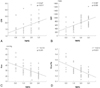
Fig. 2
The relationship between TMPG and % FDG uptake. There was a significant correlation between TMPG and % FDG uptake. FDG: 18F-fluorodeoxyglucose, TMPG: TIMI myocardial perfusion grade.
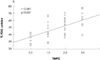
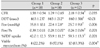
Fig. 3
Comparison of left ventricular function between at admission and at follow-up. There was a significant improvement of EF (A) in the group 3 and a significant improvement of RWMA (B) in the group 2, 3. EF: ejection fraction, RWMA: regional wall motion abnormality.
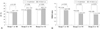
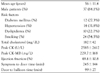
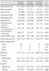
Table 3
Comparison of coronary hemodynamic results and myocardial viability between three groups after primary PCI
*p<0.05, group 1 vs group 2, †p<0.05, group 1 vs group 3, by oneway ANOVA, Bonferroni, ‡Between three groups, by Pearson's chi-square test. PCI: percutaneous coronary intervention, CFR: coronary flow reserve, DDT: diastolic deceleration time, Pcw: coronary wedge pressure, Pa: mean aortic pressure, Pcw/Pa: coronary wedge pressure/mean aortic pressure, FDG: 18F-fluorodeoxyglucose
References
1. Braunwald E. Myocardial reperfusion, limitation of infarct size, reduction of left ventricular dysfunction and improved survival: should the paradigm be expended? Circulation. 1989. 79:441–444.
2. Linderer T, Guhl B, Speilberg C, Wunderlich W, Schnitzer L, Schroder R. Effect on global and regional left ventricular functions by percutaneous transluminal coronary angioplasty in the chronic stage after myocardial infarction. Am J Cardiol. 1992. 69:997–1002.
3. Gibson CM, Cannon CP, Murphy SA, et al. Relationship of TIMI myocardial perfusion grade to mortality after administration of thrombolytic drugs. Circulation. 2000. 101:125–130.
4. Stone GW, Peterson MA, Lansky AJ, Dangas G, Mehran R, Leon MB. Impact of normalized myocardial perfusion after successful angioplasty in acute myocardial infarction. J Am Coll Cardiol. 2002. 39:591–597.
5. Ito H, Maruyama A, Iwakura K, et al. Clinical implication of the 'no reflow' phenomenon: a predictor of complications and left ventricular remodeling in reperfused anterior wall myocardial infarction. Circulation. 1996. 93:223–228.
6. Ito H, Iwakura K. Assessing the relation between coronary reflow and myocardial reflow. Am J Cardiol. 1998. 81:Suppl 12A. 8G–12G.
7. Henriques JP, Zijlstra F, Ottervanger JP, et al. Incidence and clinical significance of distal embolization during primary angioplasty for acute myocardial infarction. Eur Heart J. 2002. 23:1112–1117.
8. Ragosta M, Camarano G, Kaul S, Powers ER, Sarembock IJ, Gimple LW. Micorvascular integrity indicates myocellular viability in patients with recent myocardial infarction: new insights using myocardial contrast echocardiography. Circulation. 1994. 89:2562–2569.
9. Mazur W, Bitar JN, Lechin M, et al. Coronary flow reserve may predict myocardial recovery after myocardial infarction in patients with TIMI grade 3 flow. Am Heart J. 1998. 136:335–344.
10. Teiger E, Garot J, Aptecar E, et al. Coronary blood flow reserve and wall motion recovery in patients undergoing angioplasty for myocardial infarction. Eur Heart J. 1999. 20:285–292.
11. Lepper W, Hoffmann R, Kamp O, et al. Assessment of myocardial reperfusion by intravenous myocardial contrast echocardiography and coronary flow reserve after primary transluminal coronary angiography in patients with acute myocardial infarction. Circulation. 2000. 101:2368–2374.
12. Yoon MH, Tahk SJ, Choi SY, et al. Comparison between microvascular integrity indexes assessed by pressure/Doppler wire and %FDG uptake in AMI following primary PCI. Korean Circ J. 2006. 36:701–709.
13. Yoon MH, Tahk SJ, Choi SY, et al. Coronary flow reserve as a predictor of long-term clinical outcome after acute myocardial infarction. Korean Circ J. 2002. 32:756–765.
14. Choi SY, Tahk SJ, Yoon MH, et al. Comparison of TIMI myocardial perfusion grade with coronary flow reserve for prediction of recovery of LV function and LV remodeling in acute myocardial infarction. Korean Circ J. 2004. 34:247–257.
15. Yamamuro A, Ito H, Iwakura K, et al. Pressure-derived collateral flow index as a parameter of microvascular dysfunction in acute myocardial infarction. J Am Coll Cardiol. 2001. 38:1383–1389.
16. Shimada K, Saknoue Y, Kobayashi Y, et al. Assessment of myocardial viability using coronary zero flow pressure after successful angioplasty in patients acute anterior myocardial infarction. Heart. 2003. 89:71–76.
17. Balachandran KP, Berry C, Norrie J, et al. Relation between coronary pressure derived collateral flow, myocardial perfusion grade, and outcome in left ventricular function after rescue percutaneous coronary intervention. Heart. 2004. 90:1450–1454.
18. Tamaki N, Kawamto M, Takahashi N, et al. Prognostic value of increase in fluorine-18 deoxyglucose uptake in patients with myocardial infarction: comparison with stress thallium imaging. J Am Coll Cardiol. 1993. 22:1621–1627.
19. Sawada SG, Allman KC, Muzik O, et al. Positron emission tomography detects evidence of viability in rest technitium-99m sestamibi defects. J Am Coll Cardiol. 1994. 23:92–98.
20. Hachamovitch R, Berman DS, Shaw LJ, et al. Incremental prognostic value of myocardial perfusion single photon emission computed tomography for the prediction of cardiac death: differential stratification for risk of cardiac death and myocardial infarction. Circulation. 1998. 97:535–543.
21. De Sutter J, De Winter F, van de Wiele C, De Bondt P, D'Asseler Y, Dierckx P. Cardiac fluorine-18 fluorodeoxyglucose imaging using a dual-head gamma camera with coincidence detection: a clinical pilot study. Eur J Nucl Med. 2000. 27:676–685.
22. Segall G. Assessment of myocardial viability by positron emission tomography. Nucl Med Commun. 2002. 23:323–330.
23. Schiller NB, Shah PM, Crawford M, et al. Recommendations for quantitation of the left ventricle by two-dimensional echocardiography. J Am Soc Echocardiogr. 1989. 2:358–367.
24. Slart RH, Bax JJ, van Veldhuisen DJ, et al. Prediction of functional recovery after revascularization in patients with coronary artery disease and left ventricular dysfunction by gated FDG-PET. J Nucl Cardiol. 2006. 13:210–219.
25. Akasaka T, Yoshida K, Kawamoto T, et al. Relation of phasic coronary flow velocity characteristics with TIMI perfusion grade and myocardial recovery after primary percutaneous transluminal angioplasty and rescue stenting. Circulation. 2000. 101:2361–2367.
26. Hoffmann R, Haager P, Lepper W, Franke A, Hanrath P. Relation of coronary flow pattern to myocardial blush grade in patients with first acute myocardial infarction. Heart. 2003. 89:1147–1151.
27. Di Carli MF, Maddahi J, Rokhsar S, et al. Long-term survival of patients with coronary artery disease and left ventricular dysfunction: implications for the role of myocardial viability assessment in management decisions. J Thorac Cardiovasc Surg. 1998. 116:997–1004.
28. Yoon SN. Assessment of myocardial viability using PET. Korean J Nucl Med. 2005. 39:133–140.
29. Bacharach SL, Bax JJ, Case J, et al. PET myocardial glucose metabolism and perfusion imaging: part I. guidelines for data acquisition and patient preparation. J Nucl Cardiol. 2003. 10:543–556.
30. Schöder H, Campisi R, Ohtake T, et al. Blood flow-metabolism imaging with positron emission tomography in patients with diabetes mellitus for the assessment of reversible left ventricular contractile dysfunction. J Am Coll Cardiol. 1999. 33:1328–1337.
 XML Download