

 PDF
PDF ePub
ePub Citation
 Print
Print


Introduction
Drug-eluting stents(DESs) are commonly used in daily practice during percutaneous coronary intervention(PCI). Several significant complications after DES implantations have been recently reported. Among them, stent thrombosis(ST) is a rare and serious complication. A stent fracture is also a very rare event after a DES implantation. We report here on a case of very late stent thrombosis that occurred in a patient 24 months after a sirolimus-eluting stent(SES) implantation, and this might have been related to a stent fracture.
Case
A 58-year old male was admitted with an inferior ST segment elevation myocardial infarction(STEMI). He had been transferred to our hospital due to 4 hours of sustained chest pain. The initial ECG exhibited ST segment elevation in the inferior leads during sinus rhythm. Two years prior to his admission, he had undergone two stent implantations to the right coronary artery(RCA) because of an inferior STEMI.
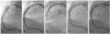
Two years previously, he was transferred to our hospital for the management of an inferior wall STEMI after thrombolytic therapy had been performed for treating the inferior STEMI. As the cardiovascular risk factors, he had a history of hypertension and hyperlipidemia and he was a current smoker. The coronary angiogram revealed two focal stenotic lesions in the proximal and mid RCA(Fig. 1A). After predilation with a 3.0 mm balloon, a 3.5×23 mm SES (Cypher, Cordis, Miami Lakes, Florida, USA) was implanted at 16 atm in the mid RCA, and a 4.0×18 mm BX-Velocity(Cypher, Cordis, Miami Lakes, Florida, USA) was implanted at 16 atm, without overlapping, in the proximal RCA after predilation with a 3.0 mm balloon. There were no events during the 8 month clinical follow-up. He underwent a follow-up coronary angiogram 8 months later as part of a workup for noncardiac surgery. There was minimal neointimal hyperplasia around both stents(Fig. 1B). Two months later, his antiplatelet therapy was discontinued for performing a left nephrectomy that was due to multiple renal stones and obstructive uropathy. After the operation, he received aspirin 100 mg daily as antiplatelet therapy. The 14 month clinical follow-up after the operation was uneventful.
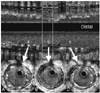
Primary PCI was then performed. The angiogram revealed total occlusion of the mid RCA at the previous SES implantation site(Fig. 1C). A floppy wire was easily passed. A large amount of thrombi was noted after predilation with a 2.5 mm balloon. After repeated 3.5 mm ballooning, intravascular ultrasound was performed to obtain more information on the occluded site. During the automatic pull-back, a sudden loss of a stent strut was noted. There was a stent fracture in the mid portion of the SES and also minimal neointimal hyperplasia(Fig. 2). Because we had already obtained optimal results and there was minimal proliferation of the neointima, we did not perform any additional stent implantations(Fig. 1D). The patient was discharged with taking a triple antiplatelet medication regimen(aspirin 200 mg, clopidogrel 75 mg, cilostazol 200 mg). There were no adverse cardiac events noted during the 9 months clinical follow-up. A second follow-up coronary angiography was performed 7 months after the stent thrombosis had occurred; the angiography did not show any evidence of restenosis(Fig. 1E).
Discussion
The DES has revolutionized the treatment of coronary artery disease, and the randomized studies have show that DES has brought about a significant reduction in the need for repeated revascularizations and a reduced rate of major adverse cardiac event.1) However, concerns have been raised about the safety of those stents in relation to the occurrence of stent thrombosis.2-4) ST with DESs may be associated with significantly higher mortality rates that are related to myocardial infarctions. This rare, but serious complication has drawn the attention of interventional cardiologists. A large portion of STs occur acutely(within 24 hours after the index procedure) or subacutely(between 1 and 30 days after).5) However, the focus of concern for the STs was that no end point for late stent thrombosis(LAST: 30 days after) could be observed, even though the incidence of this complication was small. A recent report showed significantly higher rates of death and myocardial infarctions between 6 to 18 months after the procedure in the DES group compared to the BMS group(4.9% in the DES group vs. 1.3% in the BMS group; p=0.01).4) This astonishing result was interpreted as the clinical consequence of late thrombotic events in the DES implanted patients. ST is mostly related to the discontinuation of the antiplatelet therapy.6) However, several different additional factors such as a delayed endothelialization,7) chronic inflammation or hypersensitivity to the polymer,8)9) late stent malappositioning,7)10)11) and stent underexpansion12) also affect this serious complication. Most of the mechanisms are hypothetical, and especially those that are concerned with very late stent thrombosis, and so more experience and data are needed to understand this phenomenon.
Stent fractures have recently been suggested as a new mechanism of the adverse cardiac events after DES implantation. SES fractures have also been observed by interventional cardiologists. According to the reports, stent fractures might occur with a very tortuous or greatly moved vessels that induce excessive mechanical stress during the contractions and flexion of the heart, heavy calcified lesions, long stents, overlapping sites of the stents, larger-sized ballooning sites and especially balloonmg with high pressures.13) Several cases have been reported in which the stent fractures might have been related to the restenosis after the SES implantation.13)14) However, the data regarding stent fractures and restenosis has been very limited up to the present. We experienced eight cases with SES stent fractures, and three of these cases experienced adverse cardiac events. Because of the uncertainty of the related mechanism between stent fractures and adverse cardiac events, delicate attention should be paid to the fracture site.
The case we report here suggests the existence of a new potential mechanism for very late stent thrombosis (occurring more than 1 year after the procedure) after the implantation of an SES. Stent fractures might induce mechanical injury to the endothelium, which could produce a stent thrombosis. Because our patient's fracture developed in the late period of the clinical follow-up, the antiplatelet therapy may have been insufficient to prevent thrombosis formation. Therefore, longer dual antiplatelet therapy may be needed for the groups of patients with a high risk for stent fractures.
Because no IVUS examination was performed during the first follow-up coronary angiogram, the stent fracture may have existed prior to that event. If so, the two events may have developed simultaneously apart from each other. Another possible mechanism of this event is that chronic inflammation, which developed at the fracture site, might have induced atherosclerotic plaque growth and rupture. However, there was minimal intimal hyperplasia observed on the follow-up angiogram. The possibility of plaque formation at the fracture site after the follow-up angiogram was slight. Even though the fracture had previously existed, thrombotic occlusion developed at the fracture site. The stent fracture may have affected or caused some part of the very late stent thrombosis in this patient. However, further experience is required to understand the definite mechanism.
In conclusion, we would like to suggest a new possible mechanism for very late stent thrombosis associated with a stent fracture. In the era of DESs, several special conditions might exist such as the possible need for a longer course of dual antiplatelet therapy than the usual recommended course for the patient groups with a high risk for stent thromboses or stent fractures. Careful selection of the stent length, type and positioning may also be required for special coronary situations. Further studies and collection of more data should be done to clarify whether this is an important issue or only a sporadic observation.
 XML Download