

 PDF
PDF ePub
ePub Citation
 Print
Print


Introduction
Pulmonary atresia with ventricular septal defect (VSD) and major aorto-pulmonary collateral arteries (MAPCAs) is still one of most difficult diseases to treat in pediatric cardiology.1-3) So far, various surgical techniques have been attempted, but the long-term prognosis at most medical centers has not been satisfactory.4-6) Staged operations, as initially proposed by the McCartney group, have not proved to be satisfactory.7-9) Other reports have suggested performing one-stage total correction,10-12) but this method has incurred high surgical mortality at our hospital.
In 1999 we devised a management strategy at our hospital to treat pulmonary atresia with VSD and MAPCAs, and this strategy was the result of a series of surgical attempts with using various techniques. The strategy focuses on promoting the growth of the pulmonary arterial confluence(PAC), if small, as early as possible. The flow through the PAC is augmented by the right ventricle via a pulmonary artery conduit operation(RV-PA conduit), and later by balloon and surgical angioplasty.13) Unifocalization is delayed unless absolutely necessary.14) If the PAC was absent or large, then the management plan was discussed among the surgeons after the patient underwent a complete work-up. This strategy was applied to patients who were consecutively diagnosed with pulmonary atresia with VSD and MA PCAs since the year 2000. This study was undertaken to review our surgical results and the mid-term outcomes since our treatment policy changed in 2000. The first four cases were the subjects of a previous preliminary report.15)
Subjects and Methods
The study subject consisted of all the consecutive cases seen at Seoul National University Children's Hospital from January, 2000 to December, 2003. The criteria for inclusion were 1) pulmonary atresia with VSD and MAPCAs in the normal left ventriculo-aortic connection, and 2) those patients who had not had any prior interventions. Those patients who had a patent ductus arteriosus were excluded, as were those patients with an abnormal ventriculo-arterial connection.
The hospital records, echocardiograms, catheterization data, angiocardiograms and surgical records were reviewed to collect 1) the demographic findings, 2) the anatomic details of the disease, 3) the surgical methods and 4) the long-term outcomes. The size of the branch pulmonary artery was documented in the operation records. If these records were unavailable, then the angiocardiograms and echocardigrams were used to measure the right and left branch pulmonary arteries. The size of the pulmonary artery confluence was(the diameter of the right pulmonary artery+the diameter of the left pulmonary artery)/2.
After performing palliative procedures, closure of the VSD was attempted when all or most of the PA branches were connected to the PAC and when the pressure of the right ventricle(RV) was expected to be normal or slightly elevated after the surgical closure of the VSD. When the pressure of the RV was too high after the closure of the VSD in the surgical field, then a fenestration was made in the patch, which is a subtotal correction. The palliated patients were those who were determined to be unsuitable for VSD closure.
Statistical analysis was not performed because of the small number of cases.
Results
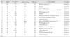
The demographic findings are shown in Table 1. There were 17 cases, among which 10 were male and 7 were female. The age at the time of diagnosis ranged from 8 days to 34 months with a median of 3 months. The median age at the first operation was 5 months, and the patients' ages ranged from 17days to 37 month. With regard to a PAC, 15 cases had a PAC and the other 2 cases did not have any PAC. When a PAC was present, the diameter ranged from 1.5mm to 8 mm. There were various initial surgical approaches: one-stage total correction(4 cases), interposition of an RV-PA conduit(3 cases), conduit insertion with additional MAPCAs procedures(8 cases) and conduit interposition from the RV to the MAPCAs(2 cases). There were 3 initial surgical deaths.
We followed up all the survivors of the initial surgery and the follow-up period varied from 2.6 years to 6.4 years with a mean of 4.6 years. Two patients died during follow up period, one of whom died of redo surgery and the other, who was in a palliated state, died of right heart failure at another hospital.
Table 2 shows the long-term outcomes according to the initial surgical methods. As a whole, 7 out of the 17 study patients underwent total correction. In addition to all three surgical survivors of one-stage total correction, 4 patients underwent staged total correction. While all 3 of the patients who had had a conduit operation without any additional vascular procedure underwent total corrective operations, only 1 of the 8 patients who had had additional vascular procedures underwent total corrective operations. The additional vascular procedures seemed to have had an unfavorable effect.
Table 3 shows the long-term outcomes according to the initial PA morphology. In this study, the PA morphology was classified arbitrarily into 3 groups: no PAC, a PAC smaller than 4 mm and a PAC larger than 4 mm. Because of the small number of cases, any firm conclusions can not be drawn, but it looks as if the size of a PAC does not affect the long-term outcome.
During the follow-up, most patients required a catheter or surgical interventions. The follow-up interventions were tabulated according to the initial surgical methods and the final outcomes(Table 4). Three survivors of one-stage total correction required balloon pulmonary angioplasty(1 case), redo Rastelli(2 cases) and surgical closure of the residual VSD(1 case). Four patients who had undergone staged total correction required multiple catheters or surgical interventions before and after the total corrective operations. All those patients who are still being palliated also required multiple catheters or surgical interventions.
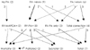
Fig. 1 shows the flow diagram of the patients. As expected, this flow diagram is quite complicated partly because of the variable nature of the disease and partly because of the different surgical options that were used. Those patients who had larger PAs were more likely to undergo total correction. Those patients who had smaller PAs were more likely to undergo RV-PA conduit with or without additional procedures such as MAPCAs unifocalization or ligation. Those patients who didn't undergo additional vascular procedures seemed more likely to undergo a total corrective operation than those patients who underwent additional vascular procedures. The most common reasons of not being able to undergo total correction were stenosis of multiple branches of the PA, anastomotic failure of the pulmonary segments, an insufficient number of unifocalized lung segments and pulmonary vascular obstructive changes.
All twelve surviving patients are being regularly checked at our outpatient clinic. All those who underwent total or subtotal correction are showing no symptoms and they are thought to be in the NYHA functional class 1. However, two patients who are still being palliated have dyspnea or cyanosis.
Discussion
Pulmonary atresia with VSD and MAPCAs can vary according to the PA anatomy and also according to the source of the pulmonary blood flow.16) Many surgical options have been devised and they have shown various success rates. The long-term prognosis of this disease is not very good and most failures are due to either an insufficient number of connected bronchopulmonary segments or to pulmonary vascular obstructive disease. One-stage total correction at an early age can prevent the development of vascular obstructive disease, but not the development of pulmonary arterial branch stenosis after surgery. Moreover, one-stage total correction entails higher surgical mortality and this procedure is very difficult to perform in some cases. The staged approach also has many drawbacks. In short, there is no surgical option that is highly successful and at the same time, universally applicable. In fact, our study showed that this is indeed the case. Various surgical options have been used despite the high surgical mortality and the poor long-term morbidity rates. However, careful analysis of our material suggests there are better surgical options depending on the PA anatomy.
In our view, one-stage total correction seems feasible when there is a large confluent PA, and this procedure has a reasonable surgical mortality rate and a low long-term morbidity rate. For those patients with a small confluent PA, the RV-PA conduit seemed to be a good option that results in total correction. However, performing additional MAPCAs procedures, in association with a conduit operation, may jeopardize the long-term prognosis. The reason why additional procedures complicate the long-term prognosis is not clear, but additional procedures are closely associated with the later development of stenosis in the pulmonary arterial tree.17)18) Simple ligation of MAPCAs is also associated with stenosis in the later stages. Moreover, this stenosis is very hard to balloon, even with performing surgical pulmonary angioplasty. Our result seems to suggest that procedures done on a small branch PA or MA PCAs are the least likely to succeed and the most likely to develop later stenosis, and so these procedures should be avoided. In contrast, simple anastomosis (RV-PA conduit) shows better results. This conduit operation may enhance growth of the pulmonary artery19)20) and a larger PA, which in turn may improve the surgical result of anastomosis between the PA and MAPCAs. After the conduit procedure, close monitoring of the pulmonary arterial tree with echocardiography and catheterization is absolutely necessary and the liberal use of catheters or surgical interventions seems to be indicated. In summary, the surgical options for this complicated disease should be individualized and vigilant surveillance for any complication is absolutely necessary for total correction at a later date.
For a patient who already has increased pulmonary flow, the construction of a RV-PA conduit may further increase the pulmonary flow and worsen the patient's heart failure. However, as discussed in a previous paper,15) the addition of another source of pulmonary flow, in theory, does not increase flow to the lungs. Nonetheless, meticulous assessment and careful decision making should be done to determine the best surgical option for a patient who has increased flow.
A major limitation of our study is that we were unable to perform statistical analysis due to the small number of cases. Because this disease is so rare and it involves anatomy that can certainly vary, it is hardly likely that there are enough patients in a single institution for conducting a meaningful statistical analysis. Thus, designing a multi-institution study seems to be necessary to shed more light on this complex disease and its proper treatment.


 XML Download