

 PDF
PDF ePub
ePub Citation
 Print
Print


Introduction
The use of metallic stents has significantly improved outcomes after percutaneous coronary intervention(PCI) leading to stent implantation in most of the one million patients who undergo this procedure annually worldwide.1)2) Drug-eluting stents are broadly available. Several studies have demonstrated the efficacy of the sirolimus-eluting stent(SES) in preventing coronary restenosis.3-5) Nevertheless, the development of in-stent restenosis is an important unresolved issue.
Cilostazol, a potent inhibitor of phosphodiesterase III, reduces smooth muscle proliferation and intimal hyperplasia after endothelial injury.6) It lowers restenosis and target lesion revascularization rates after balloon angioplasty7) and after bare metal stenting.8)9) However, few data are available about the effect of cilostazol on clinical and angiographic outcomes after SES implantation in native coronary arteries.
In this study we investigated our hypothesis that cilostazol reduces neointimal proliferation, and subsequently leads to a reduction in restenosis rates after coronary SES implantation.
Subjects and Methods
Study design
This prospective, randomized, double-blind trial compared the effectiveness of cilostazol and clopidogrel in preventing renarrowing after PCI with SES implantation in native coronary arteries. This was evaluated by quantitative coronary angiography(QCA) before and immediately after the PCI, and at the follow-up coronary angiography(CAG). Patients with stable or unstable angina or silent myocardial ischemia, who underwent elective coronary stenting at our hospital, were recruited for this study before the intervention. Written informed consent was obtained from each patient in the study.
Subjects
Patients eligible for the study were >20 years old, and had documented myocardial ischemia detected on an exercise stress electrocardiogram or thallium myocardial scan, and angiographic evidence of a de novo target lesion stenosis of >50% but <100% in a native coronary artery. Women of childbearing potential required a negative pregnancy test and made a commitment to use contraceptives during the study. Patients with multivessel coronary artery disease were eligible, and according to their eligibility, more than one lesion per patient could be randomized. Patients with stenting for a chronic total occlusion lesion were excluded, as were those with a vessel with moderate to heavy calcification or severe tortuosity, or that included side branches of >2 mm in diameter. Other exclusion criteria were a contraindication of antiplatelet agents, the use of cilostazol during the previous six months, a prior percutaneous coronary revascularization within the previous six months, stenting for unprotected left main coronary artery stenosis, primary stenting in acute myocardial infarction(MI) within the previous 24 hours, complicating congestive heart failure, a left ventricular ejection fraction of <40%, inability to follow the protocol, known bleeding disorders, thrombocytopenia (<150,000/mm3), severe hepatic or renal dysfunction (serum aspartate aminotransferase, ≥60 IU/mL; or serum creatinine, ≥2.0 mg/dL), or a major life-threatening illness.
Randomization and antiplatelet regimen
Administration of a triple regimen(100 mg aspirin daily, 75 mg clopidogrel daily, and 100 mg cilostazol twice daily) was commenced at least two days before stenting to prevent acute and subacute stent thromboses. After one month of triple oral therapy, all eligible patients were randomly assigned to receive aspirin plus clopidogrel(75 mg daily) or aspirin plus cilostazol(100 mg twice daily) according to computer-generated randomization lists, until the follow-up CAG was performed six months later.
Stent implantation procedure
The coronary angioplasty procedure was performed with the standard Judkins technique with a femoral or radial approach. Procedural success was defined as successful stenting at the desired position with a residual stenosis of <10% by visual estimate, with grade 3 flow by the classification of the Thrombolysis in Myocardial Infarction Trial, and without residual dissection. After the implantation of an SES(Cordis, a Johnson & Johnson Company, Miami, FL), adjunctive high-pressure dilatation was performed to achieve angiographic optimization, if necessary.
Clinical follow-up
All patients underwent a clinical examination as outpatients once a month after discharge. Patients were monitored at these times for the occurrence of chest pain, bleeding, rehospitalization, MI, stroke, and the need for repeated revascularization. A complete blood count and liver and renal function tests were performed before the initial CAG, one month after stenting, and at the time of follow-up CAG. Follow-up CAG was performed six months after stenting or earlier if clinically indicated. Patients who underwent CAG at four months or less and were free of restenosis were asked to return for the six-month CAG follow-up.
Quantitative coronary angiography
Baseline CAGs were performed in multiple views after the intracoronary administration of nitroglycerin. The same views were repeated after stent implantation and at late follow-up. An experienced angiographer blinded to the treatment regimen analyzed the angiographic results with a QCA analysis system(Pie Medical Imaging, Philips, Maastricht, Netherlands). The measurements were made on end-diastolic frames. Reference vessel diameter, percent diameter stenosis, lesion length, and minimal luminal diameter(MLD) were measured before and immediately after stenting, and at the time of the follow-up CAG. We determined the acute gain in MLD(MLD after stenting minus MLD before the procedure), the late loss in MLD(MLD after stenting minus MLD at follow-up angiography), and the late loss index(the average ratio of the late loss to acute gain) for each lesion. In-stent restenosis is generally defined as 50% or more of the diameter of the stent. However, in order to investigate the effects of cilostazol on the drug-eluting stent, in-stent restenosis was defined as stenosis of 30% or more of the diameter of the stent for the analysis only. We realize that in-stent restenosis of 30% is not clinically or hemodynamically meaningful in the course of atheromatous plaque progression, but we would like to emphasize again that the aim of our study was to investigate the impact of cilostazol. Target lesion revascularization was performed in patients with a stenosis of ≥70% on QCA or ≥80% by visual estimation with ischemic symptoms.
End points of the study and definitions
The primary end point was the MLD of the first lesion stented per patient, as assessed by QCA at the follow-up CAG. Secondary angiographic end points included the percentage stenosis at the target site, the rate of restenosis, and the pattern of restenosis. Events monitored at the clinical site were the occurrence of death, MI, stroke, bleeding, repeated revascularization, cerebrovascular accident, and adverse effects of clopidogrel or cilostazol necessitating withdrawal of the medication. Acute stent thrombosis was defined as thrombotic stent closure within 24 hours of stent deployment. Subacute stent thrombosis was defined as thrombotic stent closure within 30 days after the index hospitalization. Stent thrombosis was defined as angiographically confirmed occlusion of the stented segment or, in the absence of angiography, the occurrence of MI or cardiac death. MI was diagnosed when creatinine phosphokinase-MB or troponin I levels were elevated to twice the upper limit of the normal value or higher, accompanied by chest pain lasting ≥30 minutes, or when new electrocardiographic changes were seen.
Lesions ≤10 mm in length and positioned at the unscaffolded segment(i.e. articulation or gap), the body of the stent, or the proximal or distal margin(but not both) were defined as "focal" in-stent restenosis, and those >10 mm in length that extended beyond the margins of the stent(s), or had total occlusion were defined as "diffuse" pattern in-stent restenosis.
Statistical analysis
The data are expressed as mean±standard deviation. The comparison of the clinical and angiographic variables was performed with an unpaired t test for the continuous variables and a χ2 test for the categorical variables. Correlates of binary categorical variables were determined by multivariate logistic regression and correlates of continuous variables by multiple linear regression. QCA results were compared with analysis of variance for repeated measures. Statistical analysis was performed with the SPSS version 13.0 software package(SPSS, Chicago, IL). Differences were considered statistically significant at p<0.05.
Results
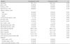
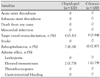
A total of 240 patients were randomly assigned to either clopidogrel(122 patients, 132 lesions) or cilostazol (118 patients, 149 lesions) after successful stent implantation between May and November 2004. Six months after stent implantation, 200 of the 240 randomized patients(83.3%) underwent quantitative coronary angiography, and were taking the study drug as directed. Both treatment groups were similar with respect to important clinical, angiographic, and procedural baseline characteristics(Table 1, 2). The target lesions of the coronary arteries, the diameters of vessels, the lesion lengths, the number of stents, and the peak deployment pressures were all similar. The two patient groups were treated similarly with respect to the stent procedure(Table 3).
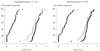
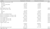
At six months, all the patients were taking the study drug as directed and underwent a follow-up QCA. Table 3 summarizes the results of QCA. Fig. 1 shows a comparison of the cumulative distribution curves of the MLDs of the cilostazol and clopidogrel groups. There were no significant differences in MLD between the two groups before or immediately after the coronary intervention, or at the follow-up angiography. Therefore, multivariable linear regression analysis failed to show that the cilostazol treatment was an independent predictor of a larger MLD(p=0.922) after we controlled for age, diabetes, hypertension, smoking, total cholesterol level, ejection fraction, lesion length, stent length, and left anterior descending artery lesion. As shown in Table 3, there were no significant differences in the restenosis rates of the cilostazol-treated and clopidogrel-treated patients(8.7% vs 11.4%, respectively, p=0.478) in the angiographic cohort. However, the in-stent restenotic lesions were shorter in the cilostazol group those in than clopidogrel group(6.26 vs 14.5 mm, respectively, p=0.001). Cilostazol treatment was associated with a focal type of in-stent restenosis pattern.
Multivariate logistic regression analysis showed that treatment with cilostazol was an independent predictor of the focal pattern of in-stent restenosis(risk ratio, 0.077; 95% CI, 0.015-0.388; p=0.02) after we controlled for age, diabetes, hypertension, smoking, total cholesterol level, ejection fraction, lesion length, stent length, and left anterior descending artery lesion. In the univariate subgroup analysis of the six-month rate of angiographic restenosis patterns(at least 30% of the reference lumen diameter), the cilostazol-treated patients who had very long lesions(more than 30 mm) experienced a highly significant(67%) reduction in the risk of a diffuse type of in-stent restenosis(p=0.011). However, other risk factors, including sex, diabetes, hypertension, smoking, stent length, left anterior descending artery site, and reference vessel diameter, showed no significant risk reduction for diffuse-type restenosis.
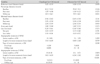
Clinical outcomes at six months were similar for the cilostazol-treated and clopidogrel-treated groups with respect to a number of important clinical events(Table 4). The occurrence rates for death, MI, stroke, bleeding, target vessel revascularization, and rehospitalization were similar in both groups. None of the patients in either group underwent emergency coronary artery bypass surgery during the follow-up period. Adverse effects of the antiplatelet drugs occurred in two patients(1.75%) in the clopidogrel group and in one patient(0.79%) in the cilostazol group(p=0.561). These involved elevated transaminase levels, but the three patients continued on the antiplatelet medication because the elevations were mild and transient.
Discussion
Coronary stents have been proven useful in compensating for the weakness of conventional balloon angioplasty, in repairing balloon-induced coronary artery dissection or abrupt vessel closure and preventing latestage restenosis. However, the limitations of coronary stenting, which include stent thrombosis and in-stent restenosis, are clinically important. A variety of factors contribute to restenosis after coronary angioplasty. These factors include inadequate dilatation, chronic elastic recoil, and neointimal proliferation.10) Coronary stenting can circumvent the problem of inadequate dilatation and minimize chronic elastic recoil, but cellular proliferation contributes greatly to in-stent restenosis.
Drug-eluting stents are now broadly available, and constituted 70% of total stent use in the recently presented registry of centers in the National Heart, Lung, and Blood Institute Dynamic Registry.11) Several studies have demonstrated the efficacy of the SES compared to that of the bare metal stent in preventing coronary in-stent restenosis.3-5) However, despite the effectiveness of drug-eluting stents in many patient subgroups, restenosis remains a significant clinical problem. Drug-eluting stents are not uniformly affordable, and this issue becomes especially problematic when multiple stents are required.13)14) Therefore, many patients remain at risk of restenosis, even in the era of drug-eluting stents, and may benefit from adjunctive systemic therapy to prevent restenosis.
This prospective, randomized, double-blinded trial investigated the effects of cilostazol on in-stent restenosis after coronary implantation of SES. The in-stent restenosis(stenosis of at lease 50% of the luminal diameter) rates of 4.4% in the clopidogrel group and 3.9% in the cilostazol group observed in this study are similar to the rate of 3.2% in SIRIUS study.3) In our study, the in-stent restenosis group was defined as those in whom at least 50% of the luminal diameter was too small to provide meaningful data about the effects of cilostazol. Therefore, we analyzed in-stent restenosis of at least 30% of the luminal diameter. More focal in-stent restenotic lesions(stenosis of at least 30% of the luminal diameter) occurred at six months in the cilostazol group than in the clopidogrel group because the restenotic lesions were significantly shorter(6.26 mm vs 14.5 mm, respectively, p=0.001), as determined by QCA, so the pattern of in-stent restenosis was focal-type in the cilostazol group(100% vs 23.1% in the clopidogrel group, p<0.001). In-stent restenosis presents in various angiographic patterns, which provide important prognostic information.15)
Cilostazol is an antiplatelet drug16) that has been widely used clinically. It has a potent inhibitory effect on platelet aggregation both in vitro and in vivo.16)17) The mechanism of the antiplatelet action of cilostazol appears to be mainly related to its inhibition of phosphodiesterase activity in the platelet cytoplasm.18) Experimental animal and human studies have shown cilostazol-associated suppression of vascular smooth muscle cell proliferation.19)20) This study does not indicate how cilostazol reduced in-stent restenosis. However, the cell activation and cell-to-cell interaction of platelets and leukocytes, which are mediated by adhesion molecules, are considered important in the mechanism of smooth muscle cell proliferation after coronary angioplasty.21-25) Coronary stenting induces the release of an adhesion molecule, P-selectin, from theα-granules of activated platelets.21)22)25) P-selectin-mediated platelet-leukocyte interactions play a crucial role in the development of stent restenosis.22)25) Cilostazol is known to inhibit the release of P-selectin by α-granules in platelets stimulated in vitro.26) In a study of 70 patients, cilostazol inhibited stent-induced P-selectin expression in platelets and the upregulation of leukocyte MAC-1. Both these events correlated with the degree of late lumen loss after coronary stent implantation.27) We speculate that inhibition of P-selectin release is one of the possible mechanisms by which cilostazol reduces stent restenosis. Cilostazol may also directly act to inhibit intimal hyperplasia. Takahashi et al.28) reported that cilostazol inhibits the incorporation of [3H] thymidine into DNA and inhibits cell growth in cultures of rat aortic smooth muscle cells stimulated with platelet-derived growth factor, insulin, and insulin-like growth factor. These actions seem to result from the inhibition of phosphodiesterase in the smooth muscle cells. In this study, the effects of therapy with cilostazol in reducing angiographic restenosis rates were rather disappointing, despite its theoretical advantages and encouraging experimental evidence.
Patients randomly assigned to treatment with cilostazol did not experience increased bleeding compared to those treated with clopidogrel or the concomitant use of clopidogrel for 30 days and aspirin for the duration of the study. Similarly, there were no differences in the rates of serious adverse cardiac events, stroke, or rehospitalization at the six-month follow-up.
The significant reduction in the lengths of the instent restenotic lesions observed in this study suggests that a reduction in neointimal hyperplasia occurred in the cilostazol-treated patients with coronary SES implantation. Our results suggest that morphological in-stent restenosis patterns can be pharmacologically modified by cilostazol. Further investigations are required to ascertain the effects of cilostazol on binary restenosis in SES implantation.
This study had several potential limitations. Although the study was a randomized trial, the randomization was performed at a single center. Because the incidence rate of stent restenosis is low, a larger cohort of patients from multiple centers is necessary to provide a more meaningful difference between the two antiplatelet regimens. On the other hand, in the era of multiple and drug-eluting stents, different stent strut designs, and stents coated with drugs of different compositions, which may all affect the results unpredictably after an immediate success or at a later follow-up, our patients were randomized as a homogeneous population treated with the same type of stent.
Target lesion revascularization was performed based on visual analysis of the angiograms in this study. However, visual estimation of the percentage stenosis was based on the nomenclature of the American Heart Association. It was performed by two independent observers unaware of the patients' assigned groups, to preclude any potential source of bias.
In our study, the late loss of MLD ascertained from the QCA results was used as an index of neointimal hyperplasia after coronary stenting. However, intimal hyperplasia cannot be evaluated strictly with QCA alone, if the phenomenon of vascular remodeling is taken into consideration. For a detailed investigation of intimal hyperplasia, an intravascular ultrasound study is necessary.
Patients included in our trial were at low risk of bleeding and had previously untreated native coronary stenoses with no severe lesion complexity, acute MI presentation, or total coronary occlusion. Whether the excluded patients, especially those likely to have thrombus-associated lesions, would benefit from the combined antiplatelet, antithrombotic, and antiproliferative effects of a drug such as cilostazol is worthy of investigation.
In conclusion, there were no significant differences in MLD between the two groups before or immediately after the coronary intervention, or at the follow-up angiography. Also, there were no significant differences in the ISR rates of the cilostazol-treated and clopidogrel-treated patients in the angiographic cohort. Cilostazol, however, was not inferior to clopidogrel in terms of clinical anticoagulation effect after SES implantation, and our study suggests that cilostazol has additional antiproliferative effects after coronary SES implantation in native coronary arteries, without an increase in adverse events.
 XML Download