

 PDF
PDF ePub
ePub Citation
 Print
Print


Abstract
The purpose of this study is to analyse clinical impact of specific MRI findings in liver in patients of long-term survivors after Kasai portoenterostomy (KPE). Twenty-eight patients who were underwent KPE were followed up more than 5 years. Macro-regenerative nodule (MRN) and beaded-duct dilatation (BDD) were considered as important findings in liver MRI. The association between these findings in MRI and clinical indicator, serum bilirubin level and history of cholangitis were evaluated. Sixteen patients (57.1%) were shown MRN in liver MRI. There were 14 patients(50%) whose MRI showed BDD. Serum total and direct bilirubin were 3.6mg/dL and 1.8mg/dL respectively in positive MRN group whereas 1.4mg/dL and 0.7mg/dL in negative MRN group (p 0.427). Serum total and direct bilirubin level were 4.2mg/dL and 2.1mg/dL in patients with BDD negative group compare to 1.1mg/dL and 0.5mg/dL in BDD positive group (p 0.281). The odds ratio to have cholangitis in the patient with MRN was 2.3 and 0.53 in patient with BDD in their MRI findings. MRN in liver MRI may suggest high bilirubin level and more chance to have cholangitis, but the findings of BDD may related to low bilirubin level and less change to have cholangitis.
Figures and Tables
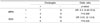
 | Fig. 1Findings of macroregenerative nodule and beaded duct appearance in MRI. A. T2-weighted MR image shows isodense nodule-like contour along the margin of the liver. B. Beaded duct dilatation which shows several high-signal small dilatations of biliary duct in T1-weighted MRI (arrows)
|
References
1. Liang JL, Cheng YF, Concejero AM, Huang TL, Chen TY, Tsang LL, et al. Macro-regenerative nodules in biliary atresia: CT/MRI findings and their pathological relations. World J Gastroenterol. 2008. 14:4529–4534.
2. Rothenberg SS, Schroter GP, Karrer FM, Lilly JR. Cholangitis after the Kasai operation for biliary atresia. J Pediatr Surg. 1989. 24:729–732.
3. Hong AR, Jung E, Kang YN, Choi SO, Park WH. Five-year Survival and Prognostic Factors after Kasai Portoenterostomy for Biliary Atresia. J Korean Surg Soc. 2010. 79:405–410.
4. Jung E, Park WH, Choi SO. Late complications and current status of long-term survivals over 10 years after Kasai portoenterostomy. J Korean Surg Soc. 2011. 81:271–275.
5. Yoon CS, Han SJ, Park YN, Chung KS, Oh Jt, Choi SH. Kasai Operation for Extrahepatic Biliary Atresia-Survival and Prognostic Factors. J Korean Assoc Pediatr Surg. 2006. 12:202–212.
6. Takahashi A, Hatakeyama S, Kuroiwa M, Suzuki N, Toki F, Suzuki M, Suehiro T, Shimura T, Kuwano H. Time-course changes in the liver of biliary atresia patients on magnetic resonance imaging. Pediatr Int. 2009. 51:66–70.
7. International Working Party. Terminology of nodular hepatocellular lesions. Hepatology. 1995. 22:983–993.
8. Liu YW, Concejero AM, Chen CL, Cheng YF, Eng HL, Huang TL, et al. Hepatic pseudotumor in long-standing biliary atresia patients undergoing liver transplantation. Liver Transpl. 2007. 13:1545–1551.
9. Takahashi A, Tsuchida Y, Suzuki N, Kuroiwa M, Ikeda H, Hirato J, et al. Incidence of intrahepatic biliary cysts in biliary atresia after hepatic portoenterostomy and associated histopathologic findings in the liver and porta hepatis at diagnosis. J Pediatr Surg. 1999. 34:1364–1368.
10. Bu LN, Chen HL, Ni YH, Peng S, Jeng YM, Lai HS, et al. Multiple intrahepatic biliary cysts in children with biliary atresia. J Pediatr Surg. 2002. 37:1183–1187.
 XML Download