Abstract
Purpose:
Epidural hematoma (EDH) in newborn is very rare, but when it occurs it is usually due to birth injury. We have evaluated the incidence and clinical features of EDH related to birth in newborn.
Methods:
We analyzed medical records of 12 newborns diagnosed with EDH at Cheil General Hospital and Women's Health Care Center from January 2000 to December 2015 retrospectively.
Results:
The incidence of EDH related to birth was 0.01%, occurring in 1 of 10,000 live births. Of the total 12 cases, 10 occurred in male and 8 in vaginal delivery. Among them, 11 infants had evidences of birth injury. Clinical presentation was nonspecific: only 1 infant had neurologic symptoms. The temporooccipital area was the most frequent location of EDH. The median size of EDH was 3.2±0.8 cm in length and 1.2±0.7 cm in depth. Mass effect accompanied with midline shift on radiologic imaging was shown in one case. Surgical drainage was needed only in one infant with neurologic symptom and mass effect on radiologic imaging, while the others were treated conservatively.
REFERENCES
1). Takagi T., Nagai R., Wakabayashi S., Mizawa I., Hayashi K. Extradural hemorrhage in the newborn as a result of birth trauma. Childs Brain. 1978. 4:306–18.

3). Akiyama Y., Moritake K., Maruyama N., Takamura M., Yamasaki T. Acute epidural hematoma related to cesarean section in a neonate with Chiari II malformation. Childs Nerv Syst. 2001. 17:290–3.

4). Negishi H., Lee Y., Itoh K., Suzuki J., Nishino M., Takada S, et al. Nonsurgical management of epidural hematoma in neonates. Pediatr Neurol. 1989. 5:253–6.

5). Halmat A., Heckly A., Adn M., Poulain P. Pathophysiology of intracranial epidural haematoma following birth. Med Hypotheses. 2006. 66:371–4.
6). Aoki N. Epidural hematoma communicating with cephalhematoma in a neonate. Neurosurgery. 1983. 13:55–7.

7). Aoki N. Epidural haematoma in the newborn infants: therapeutic consequences from the correlation between haematoma content and computed tomography features. A review. Acta Neurochir (Wien). 1990. 106:65–7.
8). Kroon E., Bok LA., Halbertsma F. Spontaneous perinatal epidural haemorrhage in a newborn. BMJ Case Rep. 2012. 2012:pii. bcr0920114735.

9). Heyman R., Heckly A., Magagi J., Pladys P., Hamlat A. Intracranial epidural hematoma in newborn infants: clinical study of 15 cases. Neurosurgery. 2005. 57:924–9.

10). Yamamoto T., Enomoto T., Nose T. Epidural hematoma associated with cephalohematoma in a neonate-case report. Neurol Med Chir (Tokyo). 1995. 35:749–52.
11). Vinchon M., Pierrat V., Tchofo PJ., Soto-Ares G., Dhellemmes P. Traumatic intracranial hemorrhage in newborns. Childs Nerv Syst. 2005. 21:1042–8.

12). Park SM., Oh KW., Kim HM. Correlation between cephalhematomas and intracranial hematomas. J Korean Soc Neo-natol. 2008. 15:160–5.
13). Ciurea AV., Kapsalaki EZ., Coman TC., Roberts JL., Robinson JS 3rd., Tascu A, et al. Supratentorial epidural hematoma of traumatic etiology in infants. Childs Nerv Syst. 2007. 23:335–41.

14). Hymel KP. Traumatic intracranial injuries can be clinically silent. J Pediatr. 2004. 144:701–2.

15). Mallet EC., Boumahni B. Neonatal extradural hematoma. Arch Pediatr. 1996. 3:608–9.
16). Mack LA., Wright K., Hirsch JH., Alvord EC., Guthrie RD., Shuman WP, et al. Intracranial hemorrhage in premature infants: accuracy of sonographic evaluation. AJR Am J Roentgenol. 1981. 137:245–50.
17). Bejar R., Curbelo V., Coen RW., Leopold G., James H., Gluck L. Diagnosis and follow-up of intraventricular and intracerebral hemorrhages by ultrasound studies of infant's brain through the fontanelles and sutures. Pediatrics. 1980. 66:661–73.

18). Babcock DS., Han BK., Weiss RG., Ryckman FC. Brain abnormalities in infants on extracorporeal membrane oxygenation: sonographic and CT findings. AJR Am J Roentgenol. 1989. 153:571–6.

19). Adcock LM., Moore PJ., Schlesinger AE., Armstrong DL. Correlation of ultrasound with post mortem neuropathologic studies in neonates. Pediatr Neurol. 1998. 19:263–71.
20). Pang D., Horton JA., Herron JM., Wilberger JE Jr., Vries JK. Nonsurgical management of extradural hematomas in children. J Neurosurg. 1983. 59:958–71.

21). Pozzati E., Tognetti F. Spontaneous healing of acute extradural hematomas: study of twenty-two cases. Neurosurgery. 1986. 18:696–700.

22). Chen TY., Wong CW., Chang CN., Lui TN., Cheng WC., Tsai MD, et al. The expectant treatment of “asymptomatic” supratentorial epidural hematomas. Neurosurgery. 1993. 32:176–9.

23). Govaert P. Clinics in developmental medicine. In: Govaert P, Linda S editors. Cranial haemorrhage in the term newborn infant. Epidural haematoma (cephalhematoma internum, internal subperiosteal bleeding). 1st ed.Cambridge: Mac Keith Press;1994. p. 34–40.
24). Ahn DH., Eom KS., Kim DW., Park JT., Moon SK., Kang SD, et al. Traumatic acute epidural hematoma in children. J Korean Neurotraumatol Soc. 2009. 5:11–5.

25). Dhellemmes P., Lejeune JP., Christiaens JL., Combelles G. Traumatic extradural hematomas in infancy and childhood. Experience with 144 cases. J Neurosurg. 1985. 62:861–4.
Fig. 1
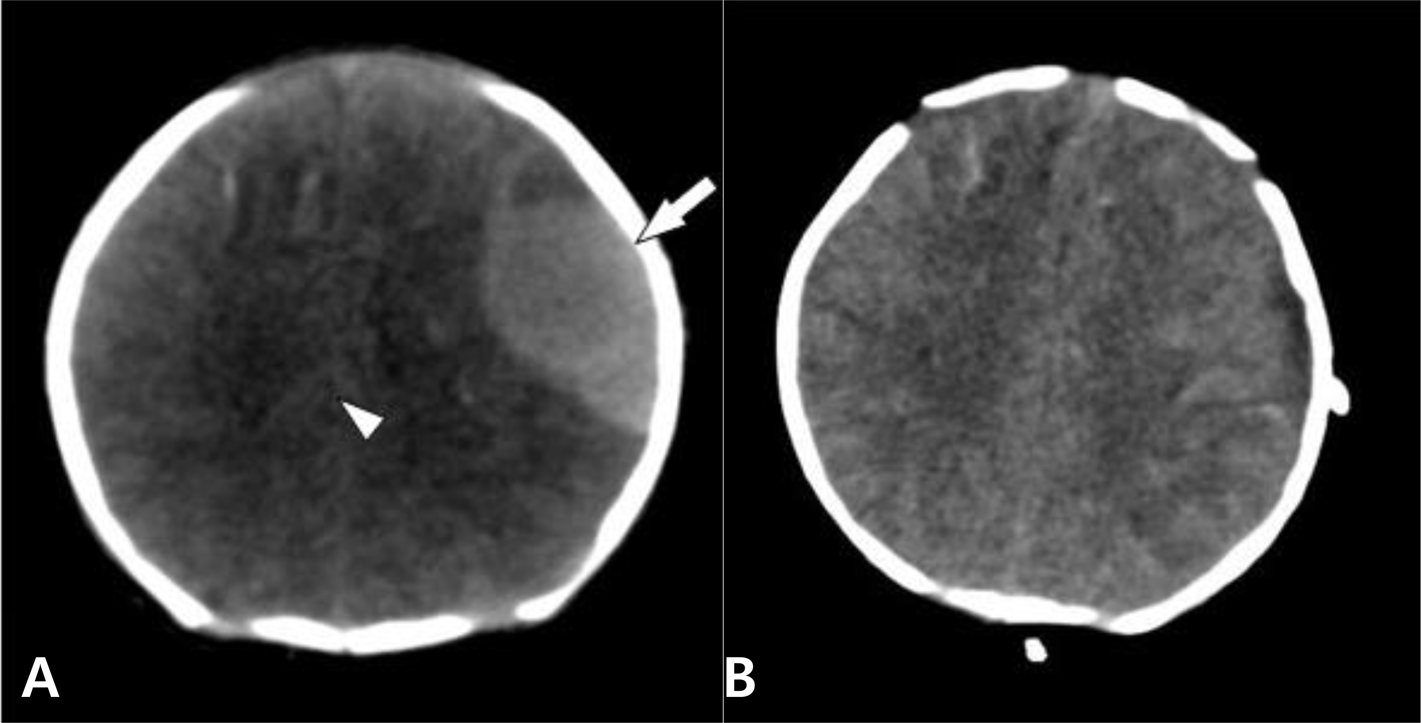
CT scans of case 10. Before (A) and after (B) surgical treatment. The EDH(arrow), shown as a lentiform high-density area on the left temporal region with mass effect(arrowhead), disappeared after surgery.

Fig. 2
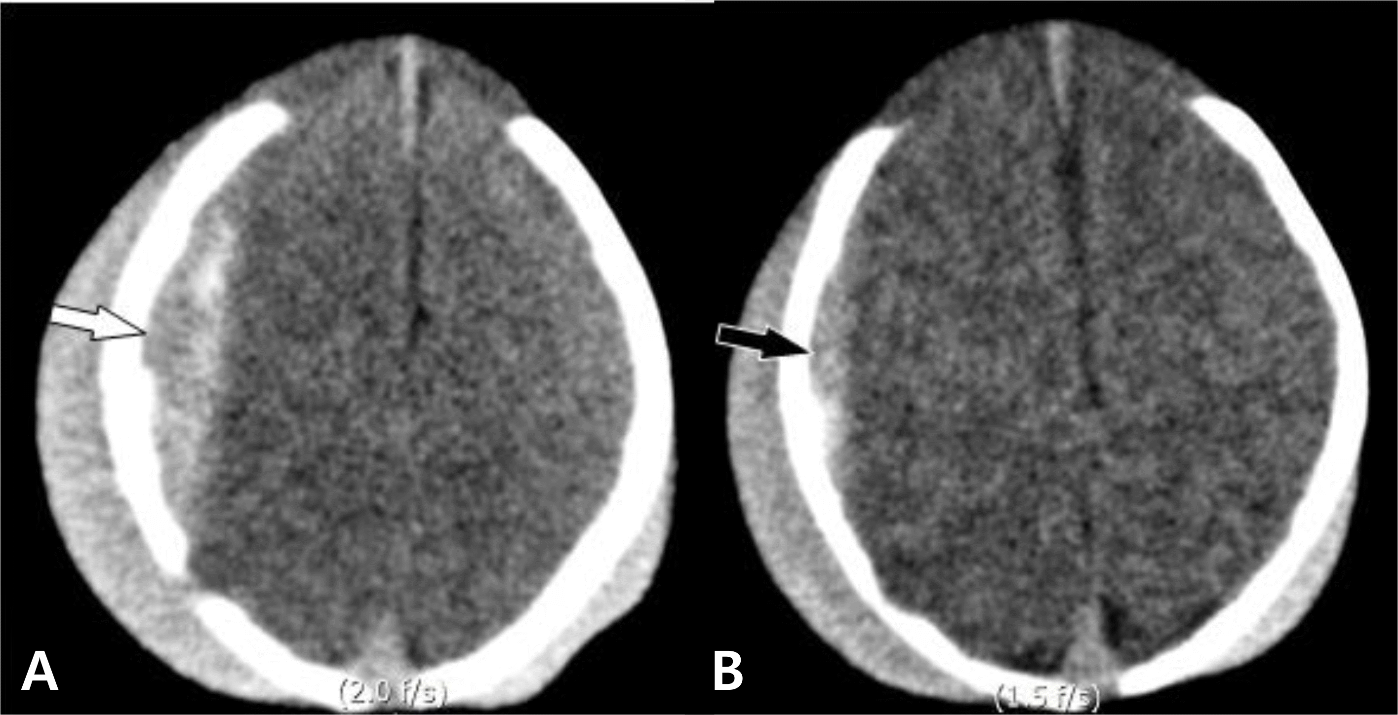
CT scans of case 12. (A) Right parietal EDH (white arrow) without mass effect was shown on day 3. (B) After 2 days, follow-up image was obtained and it showed spontaneously decreased amount of EDH (black arrow) on the same region.

Table 1.
Summary of Clinical Characteristics and Radiologic Findings of EDH


 PDF
PDF ePub
ePub Citation
 Print
Print


 XML Download