Abstract
Galloway-Mowat syndrome (GMS) is a rare autosomal recessive disorder comprising of early-onset nephrotic syndrome and central nervous system involvement including microcephaly, seizure and developmental delay. Although hiatal hernia is no longer considered essential findings for diagnosis, clinical triad of GMS included nephrotic syndrome, neurological manifestations, and hiatal hernia in the original description. We experienced a case of newborn with GMS presenting these clinical triad in neonatal period. A male infant weighing 2,250 g was born at gestational week 39+3 by cesarean section. The patient revealed mild dysmorphic facial features and microcephaly. On day 7, Nissen fundoplication was done because of hiatal hernia with gastric volvulus. At the age of 2 weeks he developed nephrotic syndrome with proteinuria and hypoalubuminemia. This is the first case of GMS that three classic findings were present in neonatal period in Korea.
References
1. Galloway WH, Mowat AP. Congenital microcephaly with hiatus hernia and nephrotic syndrome in two sibs. J Med Genet. 1968; 5:319–21.

2. Cooperstone BG, Friedman A, Kaplan BS. Galloway-Mowat syndrome of abnormal gyral patterns and glomerulopathy. Am J Med Genet. 1993; 47:250–4.

3. De Vries BB, Vant Hoff WG, Surtees RA, Winter RM. Diagnostic dilemmas in four infants with nephrotic syndrome, microcephaly and severe developmental delay. Clin Dysmorphol. 2001; 10:115–21.

4. Meyers KE, Kaplan P, Kaplan BS. Nephrotic syndrome, microcephaly, and developmental delay: three separate syndromes. Am J Med Genet. 1999; 82:257–60.

5. Jung HS, Cho EY, Lim JY, Seo JH, Choi MB, Park CH, et al. Galloway-Mowat syndrome in two siblings. J Korean Pediatr Soc. 2001; 44:1081–4.
6. Yoo BY, Cho SM, Kie KH, Jung HJ, Kim KH. A case of microcephaly and early-onset nephrotic syndrome: Galloway-Mowat syndrome. J Korean Soc Pediatr Nephrol. 2003; 7:197–203.
7. Ekstrand JJ, Friedman AL, Stafstrom CE. Galloway-Mowat syndrome: Neurologic features in two sibling pairs. Pediatr Neurol. 2012; 47:129–32.

8. Shiihara T, Kato M, Kimura T, Matsunaga A, Joh K, Hayasaka K. Microcephaly, Cerebellar atrophy, and focal segmental glomerulosclerosis in two brothers: a possible mild form of Galloway-Mowat syndrome. J Child Neurol. 2003; 18:147–9.

9. Krishnamurthy S, Rajesh NG, Ramesh A, Zenker M. Infantile nephrotic syndrome with microcephaly and global developmental delay: The Galloway-Mowat syndrome. Indian J Pediatr. 2012; 79:1087–90.
10. Dietrich A, Matejas V, Bitzan M, Hashmi S, Kiraly-Borri C, Lin SP, et al. Analysis of genes encoding laminin beta2 and related proteins in patients with Galloway-Mowat syndrome. Pediatr Nephrol. 2008; 23:1779–86.
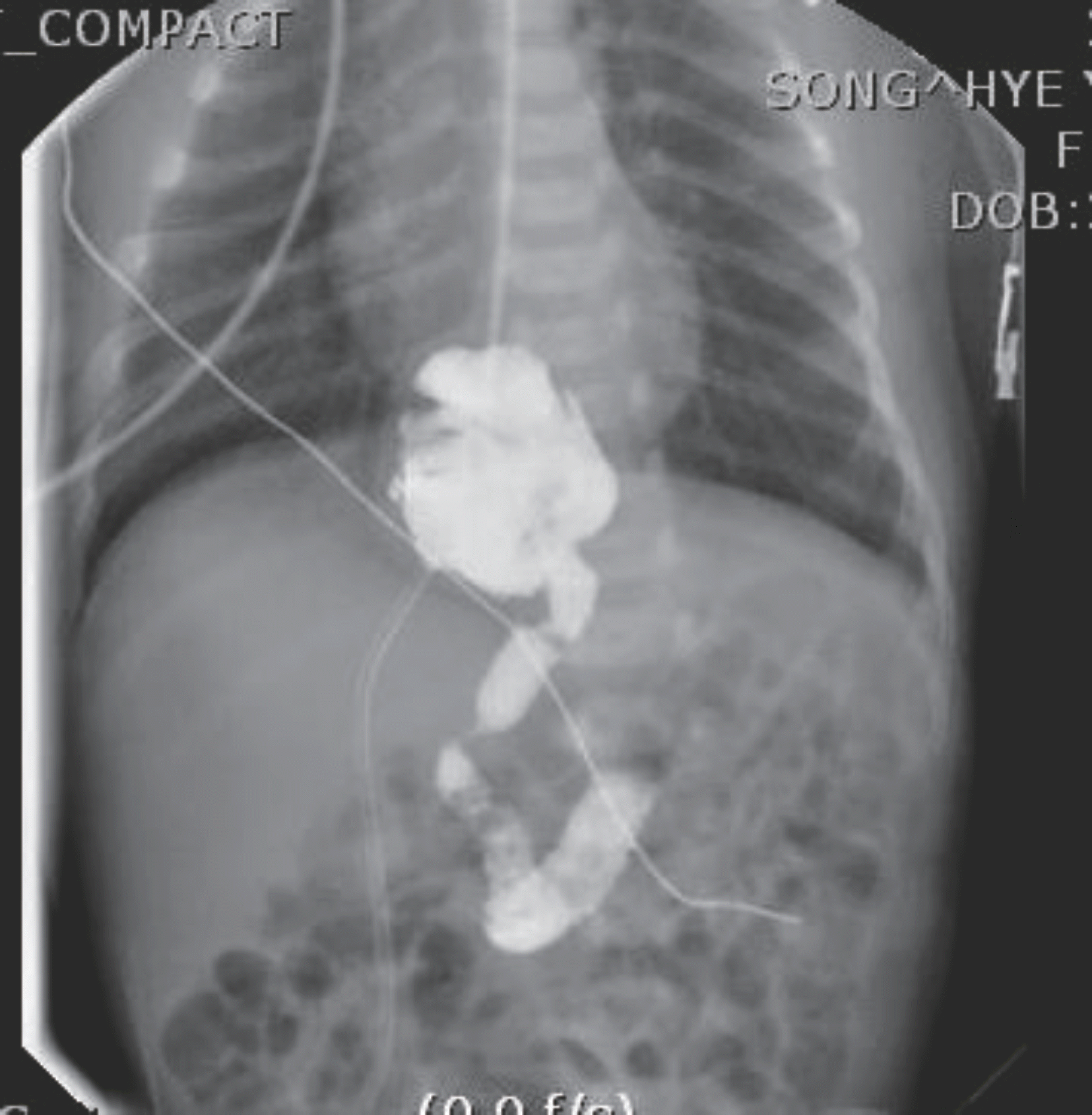
Fig. 1.
Upper gastrointestinal series show esophagogastric junction above diaphragm and folded stomach with anteriorly high located duodenal bulb.


 PDF
PDF ePub
ePub Citation
 Print
Print


 XML Download