Abstract
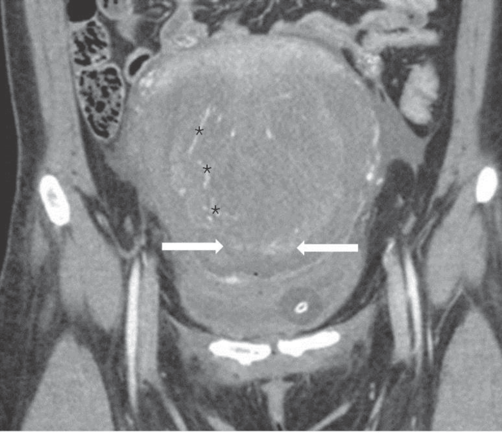
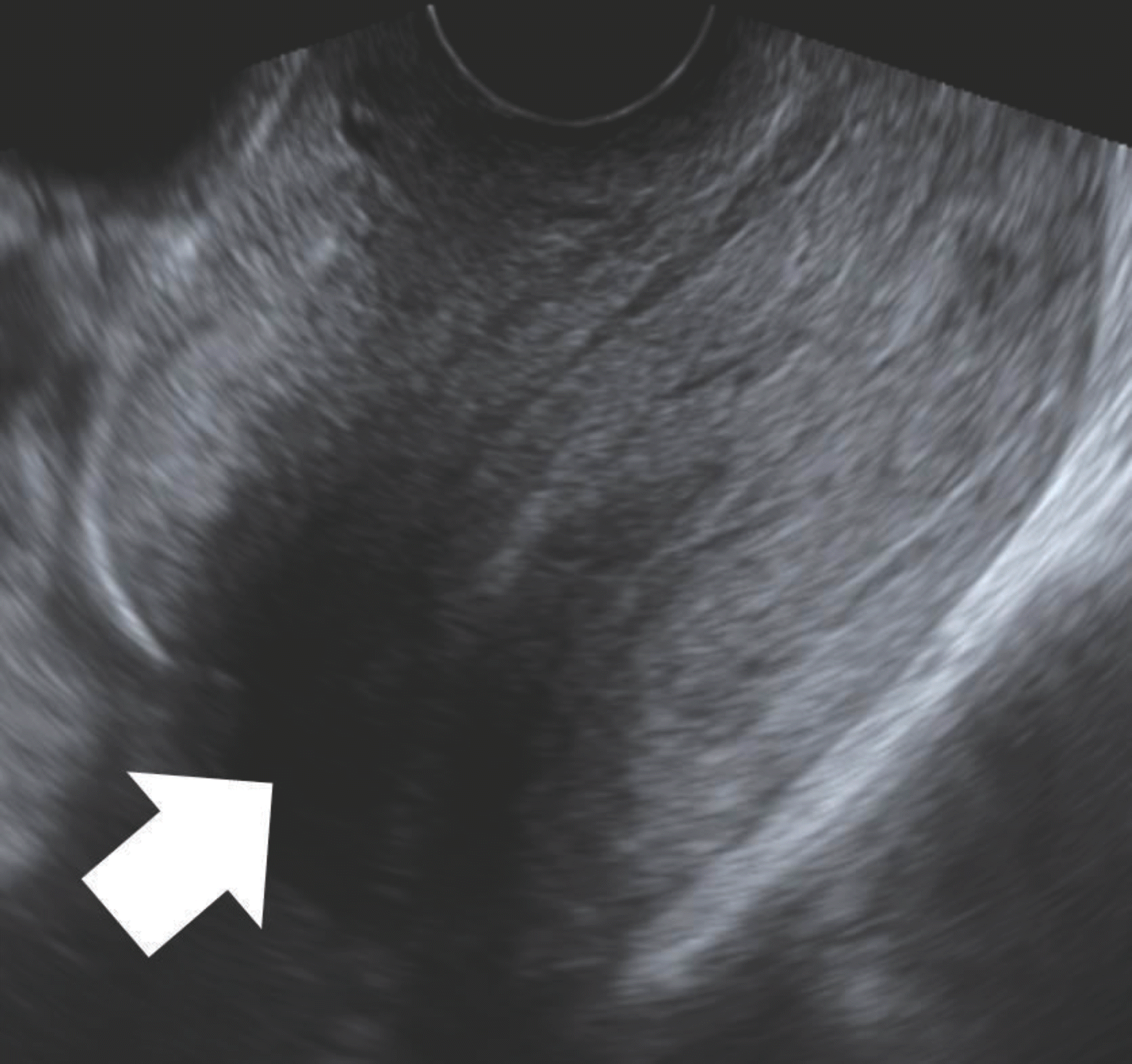
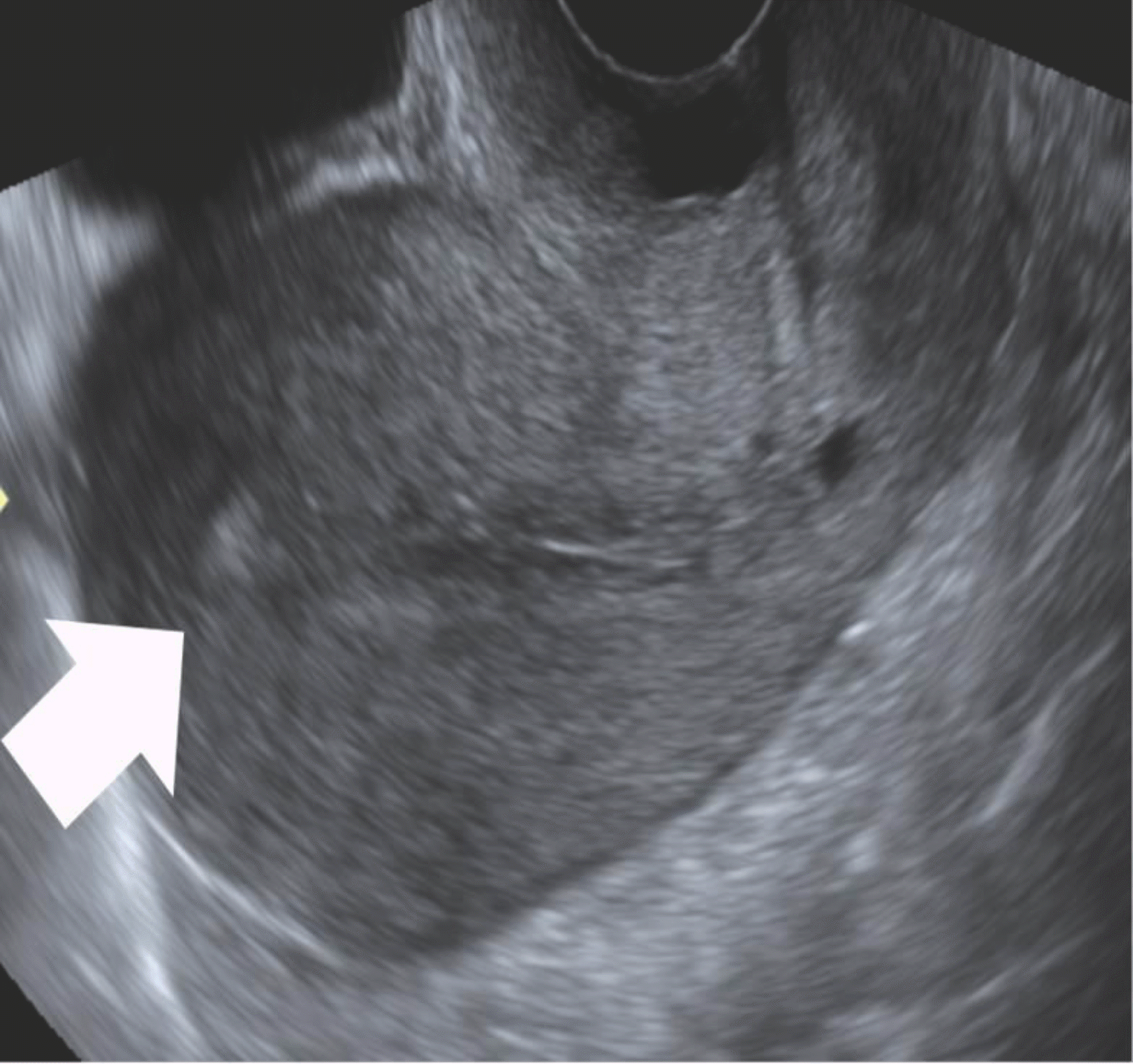
We report a case of unrecognized uterine inversion was restored spontaneously without surgical intervention. Initially, the case was diagnosed as uterine atony and not uterine inversion and was managed successfully with uterine artery embolization. However, a partial uterine inversion was detected on a subsequent scheduled pelvic examination. Fortunately, her uterus was completely restored without any surgical intervention on eighth week after delivery.
References
1. Kaya B, Tuten A, Celik H, Misirlioglu M, Unal O. Noninvasive management of acute recurrent puerperal uterine inversion with Bakri postpartum balloon. Arch Gynecol Obstet. 2014; 289:695–6.

2. Tews G, Ebner T, Yaman C, Sommergruber M, Bohau-militzky T. Acute puerperal inversion of the uterus-treatment by a new abdominal uterus preserving approach. Acta Obstet Gynecol Scand. 2001; 80:1039–40.
3. Achanna S, Mohamed Z, Krishnan M. Puerperal uterine inversion: A report of four cases. J Obstet Gynaecol Res. 2006; 32:341–5.

4. Robson S, Adair S, Bland P. A new surgical technique for dealing with uterine inversion. Aus N Z J Obstet Gynaecol. 2005; 45:250–1.

5. Tank Parikshit D, Mayadeo Niranjan M, Nandanwar YS. Pregnancy outcome after operative correction of puerperal uterine inversion. Arch Gynecol Obstet. 2004; 269:214–6.

7. Adesiyun AG. Septic postpartum uterine inversion. Singapore Med J. 2007; 48:943–5.
Fig. 1.
MDCT showed enhanced vascularity (asterisks) directed downwards along the inverted uterine musculature. The uterine fundus inverted into the level of the cervix (arrows). MDCT; multi-detector computed tomography.


 PDF
PDF ePub
ePub Citation
 Print
Print


 XML Download