

 PDF
PDF ePub
ePub Citation
 Print
Print


Abstract
Neonatal gastric perforation is a rare but fatal disease, occurred mainly in preterm infants. In general, primary surgical repair is the main treatment. To the best of our knowledge, there has been only one report of improvement of gastric perforation in neonates after percutaneous peritoneal drainage alone. We describe a case of gastric perforation in a premature extremely low-birth-weight infant girl of 25+4 weeks gestational age. We present this case to emphasize that gastric perforation may improve with percutaneous peritoneal drainage.
References
1. Jung YS, Kim SY. Neonatal spontaneous gastric perforation caused by Group B streptococcus (GBS) chorioamnionitis. Korean J Perinatol. 2007; 18:438–42.
2. Lin CM, Lee HC, Kao HA, Hung HY, Hsu CH, Yeung CY, et al. Neonatal gastric perforation: report of 15 cases and review of the literature. Pediatr Neonatol. 2008; 49:65–70.

3. Winn HN. Group B streptococcus infection in pregnancy. Clin perinatol. 2007; 34:387–92.
4. Duran R, Inan M, Vatansever U, Aladag N, Acunas B. Etiology of neonatal gastric perforations: review of 10 years'experience. Pediatr Int. 2007; 49:626–30.
5. Kawase Y, Ishii T, Arai H, Uga N. Gastrointestinal perforation in very low-birthweight infants. Pediatr Int. 2006; 48:599–603.
6. Rhim SY, Jung PM. Clinical study of neonatal gastric perforation. J Korean Assoc Pediatr Surg. 2005; 11:123–30.
7. Leone RJ Jr, Krasna IH. Spontaneous neonatal gastric perforation: is it really spontaneous? J Pediatr Surg. 2000; 35:1066–9.
8. Kellogg HG, Abelson SM, Cornwell FA. Perforation of the stomach in the newborn infant; a report of survival. J Pediatr. 1951; 39:357–62.
9. Thelander HE. Perforation of the gastrointestinal tract of the newborn infant. Am J Dis Child. 1939; 58:371–93.
10. Fischer D, Schloesser R, Buxmann H, Veldman A. Recombinant activated factor VII as a hemostatic agent in very low birth weight preterms with gastrointestinal hemorrhage and disseminated intravascular coagulation. J Pediatr Hematol Oncol. 2008; 30:337–42.
11. MillerRA.Observationonthegastricacidityduringthefirst month of life. Arch Dis Child. 1941; 16:22–30.
12. Yoon JM, Lim JW, Cheon EJ, Ko KO, Mok WK. The case of pseudocyst formation after spontaneous neonatal gastric perforation. J Korean Soc Neonatal. 2006; 13:273–7.
13. Chouteau W, Green DW. Neonatal gastric perforation. J perinatol. 2003; 23:345–7.
14. Pochaczevsky R, Bryk D. New roentgenographic signs of neonatal gastric perforation. Radiology. 1972; 102:145–7.
15. Nagaraj HS, Sandhu AS, Cook LN, Buchino JJ, Groff DB. Gastrointestinal perforation following indomethacin therapy in very low birth weight infants. J Pediatr Surg. 1981; 16:1003–7.
16. Gillesby WJ, Wheeler JR. Acute gastric dilatation. Am Surg. 1956; 22:1154–62.
17. Saracli T, Mann M, French DM, Booker CR, Scott RB. Rupture of the stomach in the newborn infant: report of three cases and review of the world literature. Clin pediatr (Phila). 1967; 6:583–8.
18. Aydın M, Zenciroğlu A, Hakan N, Erdoğan D, Okumuş N, İpek MS. Gastricperforationinanextremelylowbirth weight infant recovered with percutaneous peritoneal drainage. Turk J Pediatr. 2011; 53:467–70.
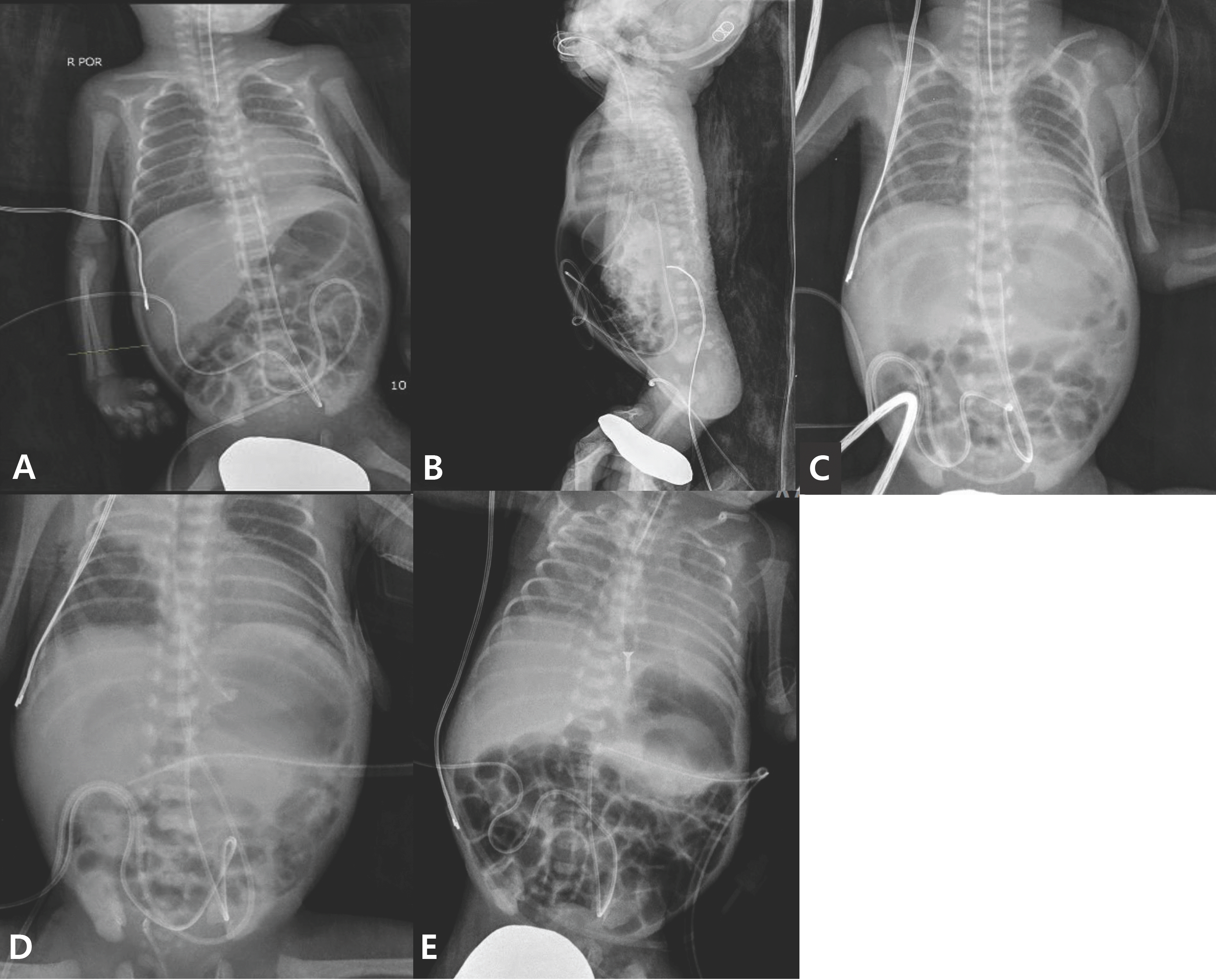
Fig. 1.
(A) On the 7th day of life, plain abdomen shows marked small bowel distension and free air in subdiaphragmatic area. (B) At the same day, a lateral abdominal X-ray shows marked free air. (C) On the 9th day of life, plain abdomen X-ray shows pneumoperitoneum. (D) Jackson-Pratt tube inserted into the peritoneum. (E) Plain abdomen on the 20th day of life. Abdominal distension is markedly decreased, and subdiaphragmatic free air is almost disappeared. Jackson-Pratt tube was removed the next day.
 XML Download