Abstract
Neonatal herpes simplex virus (HSV) encephalitis is a rare disease nowadays because of prenatal screening test and management. It shows progressive central nervous system manifestations affecting predominantly temporal and frontal lobes. Early diagnosis of HSV encephalitis is important since even with the early initiation of high-dose intravenous acyclovir therapy, it results in serious morbidity among survivors. A 14-day-old neonate with fever and poor oral intake was admitted via emergency department. The next day she had seizures and the brain was damaged with permanent sequelae despite of early administration of intravenous acyclovir on day 2 of admission. We report a serious case of HSV encephalitis diagnosed as type 2 HSV by polymerase chain reaction and culture of a newborn without proper prenatal screening test.
REFERENCES
1.Stanberry LR. Herpes Simplex Virus. Kliegman R, Behr-man RE, Jenson HB, Stanton BF, editors. editors.Nelson textbook of pediatrics. 19th ed.Philadelphia: Elsevier/Saunders;2012. p. 1097–103.

2.Brown ZA., Benedetti J., Ashley R., Burchett S., Selke S., Berry S, et al. Neonatal herpes simplex virus infection in relation to asymptomatic maternal infection at the time of labor. N Engl J Med. 1991. 324:1247–52.

3.Brown ZA., Selke S., Zeh J., Kopelman J., Maslow A., Ashley RL, et al. The acquisition of herpes simplex virus during pregnancy. N Engl J Med. 1997. 337:509–15.

4.Corey L., Wald A. Maternal and neonatal herpes simplex virus infections. N Engl J Med. 2009. 361:1376–85.

5.Lee IS. Historical changes and the present situation of sexually transmitted diseases. J Korean Med Assoc. 2008. 51:868–74.

6.Korea centers for disease control and prevention. Infectious Diseases Surveillance Yearbook, 2013. Osong: The institute;2014.
7.Park DS., Choi SD., Suh BK., Chung SY., Kang JH. A case of neonatal herpes simplex virus encephalitis. Korean J infect Dis. 1995. 27:407–12.
8.Lee B., Hwang J., Choi YH., Han YJ., Choi YH., Park JD. Disseminated neonatal herpes simplex virus infection. Korean J Crit Care Med. 2013. 28:331–5.

9.Kim T. Treatment and management of sexually transmitted disease. J Korean Med Assoc. 2008. 51:884–96.
10.Ryu KY., Hoh JK., Park MI. Preconception infection and genetic counseling. J Korean Med Assoc. 2011. 54:838–44.

11.Rudnick CM., Hoekzema GS. Neonatal herpes simplex virus infections. Am Fam Physician. 2002. 65:1138–42.
12.Jones CA., Raynes-Greenow C., Isaacs D. Neonatal HSV study investigators and contributors to the Austrailan paediatric surveillance unit. Population-based surveillance of neonatal herpes simplex virus infection in Australia, 1997– 2011. Clin Infect Dis. 2014. 59:525–31.
13.Kim ID., Chang HS., Hwang KJ. Herpes simplex virus 2 in-fection rate and necessity of screening during pregnancy: a clinical and seroepidemiologic study. Yonsei Med J. 2012. 53:401–7.

14.Kennedy PG., Steiner I. Recent issues in herpes simplex encephalitis. J. Neurovirol. 2013. 19:346–50.

15.Gilden DH., Mahalingam R., Cohrs RJ., Tyler KL. Herpesvirus infections of the nervous system. Nat Clin Pract Neurol. 2007. 3:82–94.

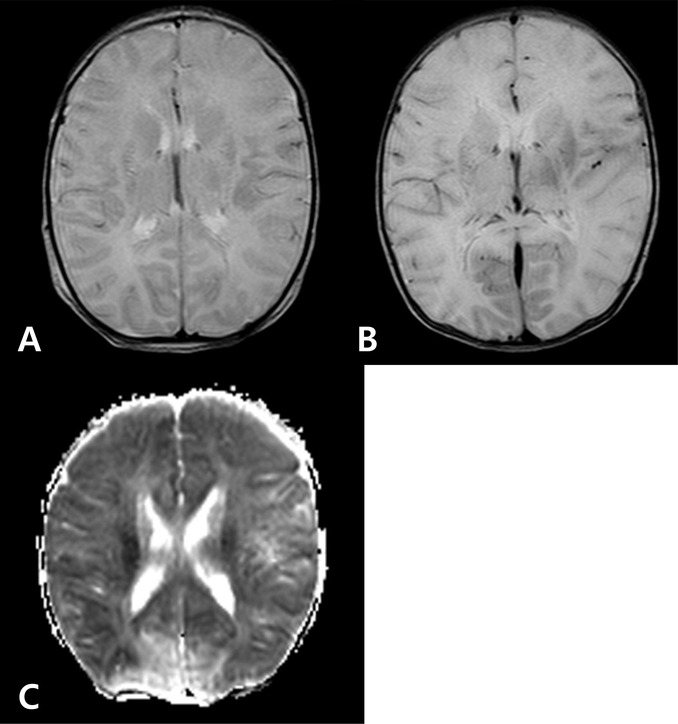
Fig. 1.
(A) T2-weighted axial images through basal ganglia on hospital day 3, showed no abnormal enhancement. (B) T2-weighted axial images through basal ganglia on hospital day 5, showed generalized increased signal. (C) Diffusion-weighted axial images through basal ganglia on hospital day 5, showed diffuse high signal intensity.

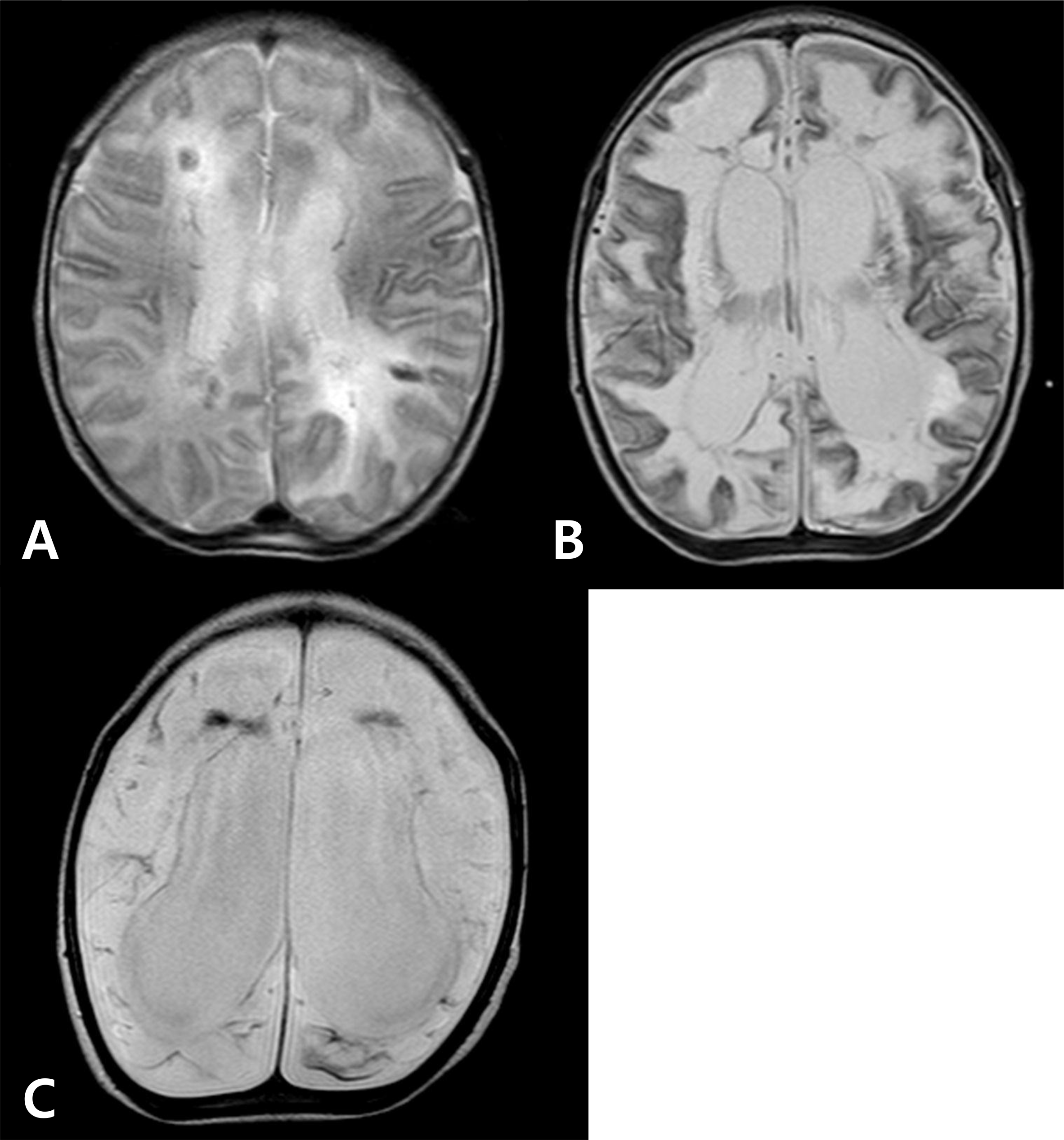
Fig. 2.
At T2-weighted axial images through basal ganglia (A) on hospital day 11, there were multifocal hemorrhages, enlarged ventricles. (B) On hospital day 26, there were marked and diffuse brain atrophy with encephalomalacia of brain. (C) Six months later after discharge, there were more progressed brain atrophy with marked encephalomalacia of entire brain.

Table 1.
Cerebrospinal fluid analysis and virus examinations


 PDF
PDF ePub
ePub Citation
 Print
Print


 XML Download