

 PDF
PDF ePub
ePub Citation
 Print
Print


Abstract
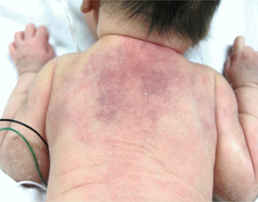
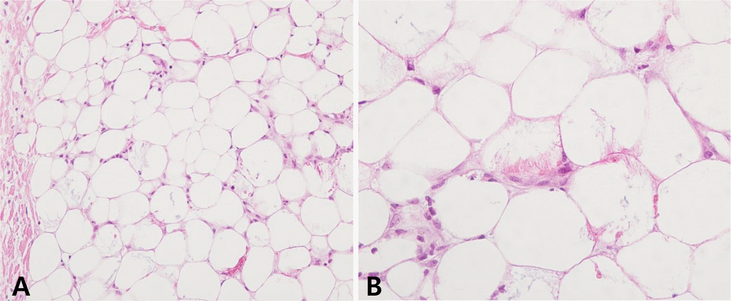
Subcutaneous fat necrosis of the newborn is a rare, benign disease usually found in full-term infants. It occurs usually in a few weeks after birth, as one or multiple indurated nodules or plaques on the fat pads-rich fraction of the body and disappeared after few weeks to months. Complications such as hypercalcemia, pain, lipid abnormalities (dyslipidemia), renal failure, and subcutaneous atrophy may occur. We report a case of subcutaneous fat necrosis associated with hypoglycemia and meconium aspiration syndrome in the term infant and review the associated literatures.
REFERENCES
1.Burden AD., Krafchik MB. Subcutaneous fat necrosis of the newborn: a review of 11 cases. Pediatr Dermatol. 1999. 16:384–7.
2.Hogeling M., Meddles K., Berk DR., Bruckner AL., Shimotake TK., Cohen RS, et al. Extensive subcutaneous fat necrosis of the newborn associated with therapeutic hypothermia. Pediatr Dermatol. 2012. 29:59–63.

3.Mitra S., Dove J., Somisetty SK. Subcutaneous fat necrosis in newborn-an unusual case and review of literature. Eur J Pediatr. 2011. 170:1107–10.
4.Bonnemains L., Rouleau S., Sing G., Bouderlique C., Coutant R. Severe neonatal hypercalcemia caused by subcutaneous fat necrosis without any apparent cutaneous lesion. Eur J Pediatr. 2008. 167:1459–61.
5.Mahé E., Girszyn N., Hadj-Rabia S., Bodemer C., Hamel-Teillac D., De Prost Y. Subcutaneous fat necrosis of the newborn: a systematic evaluation of risk factors, clinical manifestations, complications and outcome of 16 children. Br J Dermatol. 2007. 156:709–15.
6.Jeon HS., Lee MI., Ahn DH., Yoo HJ. A case of subcutaneous fat necrosis of the newborn. J Korean Pediatric Soc. 1994. 37:405–9.
7.Hong MA., Oh KC., Ahn SI., Shin HJ., Chang JK., Lee BD, et al. A case of subcutaneous fat necrosis in neonate with meconium aspiration syndrome. J Korean Pediatr Soc. 2002. 45:1422–5.
8.Choi J., Kim JS., Yoon HS., Jung EC., Lee AY., Song KY. A case of generalized subcutaneous fat necrosis of newborn. Korean J Dermatol. 2003. 41:932–5.
9.Yi KS., Cho BS., Bae IH., Lee SY., Jeon MH., Lee OJ, et al. Subcutaneous fat necrosis of the newborn: A case report. J Korean Soc Ultrasound Med. 2007. 26:125–8.
10.Zeb A., Darmstadt GL. Sclerema neonatorum: a review of nomenclature, clinical presentation, histological features, differential diagnoses and management. J Perinatol. 2008. 28:453–60.
11.Tran JT., Sheth AP. Complications of subcutaneous fat necrosis of the newborn: a case report and review of the literature. Pediatr Dermatol. 2003. 20:257–61.
12.Borgia F., De Pasquale L., Cacace C., Meo P., Guarneri C., Can-navo SP. Subcutaneous fat necrosis of the newborn: be aware of hypercalcaemia. J Paediatr Child Health. 2006. 42:316–8.
13.Oswalt GC., Montes LF., Cassady G. Subcutaneous fat necrosis of the newborn. J Cutan Pathol. 1978. 5:193–9.
14.Blake HA., Goyette EM., Lyter CS., Swan H. Subcutaneous fat necrosis complicating hypothermia. J Pediatr. 1955. 46:78–80.
15.Ivy RE., Howard FH. Subcutaneous fat necrosis of the newborn infant; report of a case in an infant born by cesarean section, and with no anoxia. J Pediatr. 1953. 42:600–2.
16.Ladoyanni E., Moss C., Brown RM., Ogboli M. Subcutaneous fat necrosis in a newborn associated with asymptomatic and uncomplicated hypercalcemia. Pediatr Dermatol. 2009. 26:217–9.
17.Akin MA., Akin L., Sarici D., Yilmaz I., Balkanli S., Kurtoglu S. Follow-up during early infancy of newborns diagnosed with subcutaneous fat necrosis. J Clin Res Pediatr Endocrinol. 2011. 3:216–8.
18.Sharata H., Postellon DC., Hashimoto K. Subcutaneous fat necrosis, hypercalcemia, and prostaglandin E. Pediatr Dermatol. 1995. 12:43–7.
19.Finne PH., Sanderud J., Aksnes L., Bratlid D., Aarskog D. Hypercalcemia with increased and unregulated 1,25-dihydro-xyvitamin D production in a neonate with subcutaneous fat necrosis. J Pediatr. 1988. 112:792–4.
 XML Download