Abstract
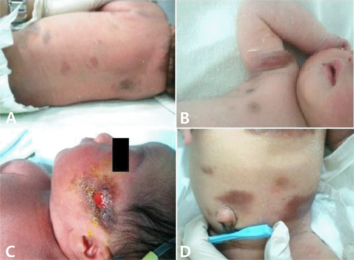
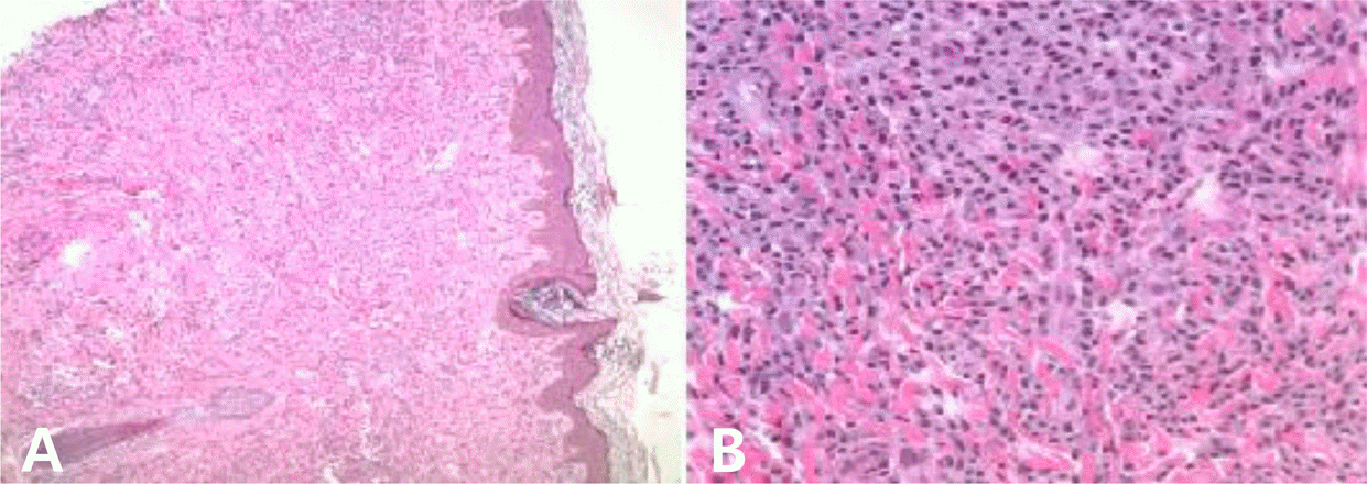
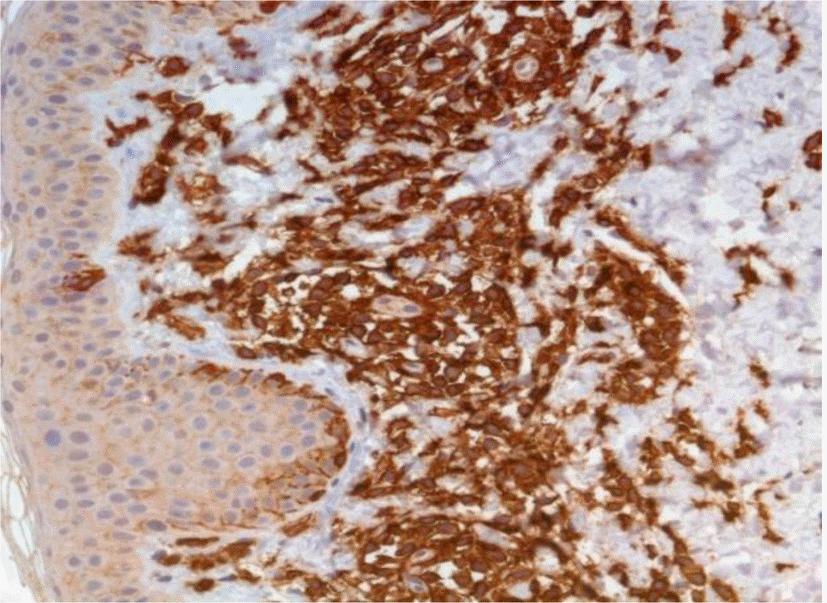
Diffuse cutaneous mastocytosis (DCM) is a rare variant of mast cell disease with widespread erythema and is clinically apparent in early infancy. We report the case of a 1-day-old female neonate who presented with diffuse flush, pruritus, and extensive blistering. DCM was diagnosed by immunohistochemical staining with anti-CD117, which revealed mast cell infiltration. DCM is a severe and heterogeneous cutaneous disease, and is associated with mast cell mediator-related symptoms and risk of anaphylactic shock. We describe this case and provide the first literature review of neonatal onset DCM in Korea.
Go to : 
REFERENCES
1.Valent P., Akin C., Escribano L., Födinger M., Hartmann K., Brockow K, et al. Standards and standardization in mastocytosis: consensus statements on diagnostics, treatment recommendations and response criteria. Eur J Clin Invest. 2007. 37:435–53.

2.Valent P., Horny HP., Escribano L., Longley BJ., Li CY., Sohwartz LB, et al. Diagnostic criteria and classification of mastocytosis: a consensus proposal. Leuk Res. 2001. 25:603–25.

3.Muñoz LS., Twose IA., Garcı´a-Montero AC., Teodosio C., Acevedo MJ., Pedreira CE, et al. Evaluation of the WHO criteria for the classification of patients with mastocytosis. Mod Pathol. 2011. 24:1157–69.

4.Koga H., Kokubo T., Akaishi M., Iida K., Korematsu S. Neonatal onset diffuse cutaneous mastocytosis: a case report and review of the literature. Pediatr Dermatol. 2011. 28:542–6.

5.Heide R., Middelkamp Hup MA., Mulder PG., Oranje AP. Clinical scoring of cutaneous mastocytosis. Acta Derm Venereol. 2001. 81:273–6.

6.Yanagihori H., Oyama N., Nakamura K., Kaneko F. C-kit mutations in patients with childhood-onset mastocytosis and genotype-phenotype correlation. J Mol Diagn. 2005. 7:252–7.

7.Chang SE., Kang SK., Jee MS., Choi JH., Sung KJ., Moon KC, et al. Clinicopathological study of 30 cases of cutaneous mastocytosis. Korean J Dermatol. 2002. 40:501–5.
8.Kanwar AJ., Dhar S. Diffuse cutaneous mastocytosis: a rare entity. Pediatr Dermatol. 1993. 10:301–2.

9.Murphy M., Walsh D., Drumm B., Watson R. Bullous mastocytosis: a fatal outcome. Pediatr Dermatol. 1999. 16:452–5.
10.Arock M., Valent P. Pathogenesis, classification and treatment of mastocytosis: state of the art in 2010 and future perspectives. Expert Rev Hematol. 2010. 3:497–516.

11.Schwartz LB., Irani AM. Serum tryptase and the laboratory diagnosis of systemic mastocytosis. Hematol Oncol Clin North Am. 2000. 14:641–57.

12.DiBacco RS., DeLeo VA. Mastocytosis and mast cell. J Am Acad Dermatol. 1982. 7:709–22.
Go to : 
 | Fig. 1.Clinical presentation: Patient at birth with diffuse erythematous rash on back (A). Partly hemorrhagic vesicle and bulla on axilla (B). Hemorrhagic crusts and erosion found on the face (C). Diffuse cutaneous mastocytosis with erythema and leathery thickened skin on abdomen (D). |


 PDF
PDF ePub
ePub Citation
 Print
Print


 XML Download