

 PDF
PDF ePub
ePub Citation
 Print
Print


Abstract
Purpose :
The aim of this study was to determine the value of a gray-level histogram of the cervix as a predictor of preterm birth in women who admitted for preterm labor.
Methods :
Ninety-seven women who admitted Chonnam national university for preterm labor were enrolled in this prospective study. A transvaginal ultrasonography for measurement of cervical length (CL), gray-scale histogram and cervical volume was performed when patients were admitted. Anterior and posterior cervical walls and AP (anterior and posterior) difference in MGL (mean gray level between anterior and posterior) were checked. And we analyzed the relationship between the value and preterm birth.
Results :
The overall rate of preterm birth before 37 weeks was 53.6% and after 37 weeks was 46.4%, respectively. Logistic regression analysis demonstrated that not only cervix length (P=0.003; odds ratio [OR], 0.189; 95% confidence interval [CI], 0.064-0.560) but also anterior histogram (P=0.028; OR, 0.319; 95% CI, 0.115-0.884) was independent predictor of preterm birth before 37 weeks. The receiver operator characteristics (ROC) curves were analyzed for the anterior histogram, a value of 85.9 was the best cut-off value to determine the preterm birth. The areas under the ROC curve indicate that the variable provides a prognostic value for the prediction for preterm birth. To predict a preterm birth, anterior histogram had 78.8% sensitivity and 46.7% specificity.
Go to : 
REFERENCES
1). Creasy RK. Preventing preterm birth. N Engl J Med. 1991. 325:727–9.

2). Goffinet F., Rozenberg P., Kayem G., Perdu M., Philippe HJ., Nisand I. The value of intravaginal ultrasonography of the cervix uteri for evaluation of the risk of premature labor. J Gynecol Obstet Biol Reprod (Paris). 1997. 26:623–9.
3). Iams JD. Prediction and early detection of preterm labor. Obstet Gynecol. 2003. 101:402–12.
4). Iams JD., Goldenberg RL., Meis PJ., Mercer BM., Moawad A., Das A, et al. The length of the cervix and the risk of spontaneous premature delivery. National Institute of Child Health and Human Development Maternal Fetal Medicine Unit Network. N Engl J Med. 1996. 334:567–72.
5). Severi FM., Bocchi C., Florio P., Picciolini E., D'Aniello G., Petraglia F. Comparison of two-dimensional and three-dimensional ultrasound in the assessment of the cervix to predict preterm delivery. Ultrasound Med Biol. 2003. 29:1261–5.
6). Maeda K., Utsu M., Yamamoto N., Serizawa M. Echogenicity of fetal lung and liver uantified by the grey-level histogram width. Ultrasound Med Biol. 1999. 25:201–8.
7). Maeda K., Utsu M., Kihaile PE. Quantification of sonographic echogenicity with grey-level histogram width: a clinical tissue characterization. Ultrasound Med Biol. 1998. 24:225–34.
8). Tekesin I., Hellmeyer L., Heller G., Romer A., Kuhnert M., Schmidt S. Evaluation of quantitative ultrasound tissue characterization of the cervix and cervical length in the prediction of premature delivery for patients with spontaneous preterm labor. Am J Obstet Gynecol. 2003. 189:532–9.
9). Kim YH., Kim JW., Kim CH., Cho MK., Cho HY., Cho AR, et al. Evaluation of length, volume and gray-scale histogram of the cervix as predictors of successful induction. Korean J Obstet Gynecol. 2010. 53:389–95.
10). Kuwata T., Matsubara S., Taniguchi N., Ohkuchi A., Ohkusa T., Suzuki M. A novel method for evaluating uterine cervical consistency using vaginal ultrasound gray-level histogram. J Perinat Med. 2010. 38:491–4.
11). Yu SY., Tozzi CA., Baviarz J., Leppert PC. Collagen changes in rat cervix in pregnancy–polarized light microscopic and electron microscopic studies. Proc Soc Exp Biol Med. 1995. 209:360–8.
12). Guzman ER., Walters C., Ananth CV., O'Reilly-Green C., Benito CW., Palermo A, et al. A comparison of sonographic cervical parameters in predicting spontaneous preterm birth in high-risk singleton gestations. Ultrasound Obstet Gynecol. 2001. 18:204–10.
13). Park IY., Kwon JY., Kwon JY., Hong SC., Choi HM., Kwon HS, et al. Usefulness of cervical volume by three-dimensional ultrasound in identifying the risk for preterm birth. Ultrasound Med Biol. 2011. 37:1039–45.
Go to :
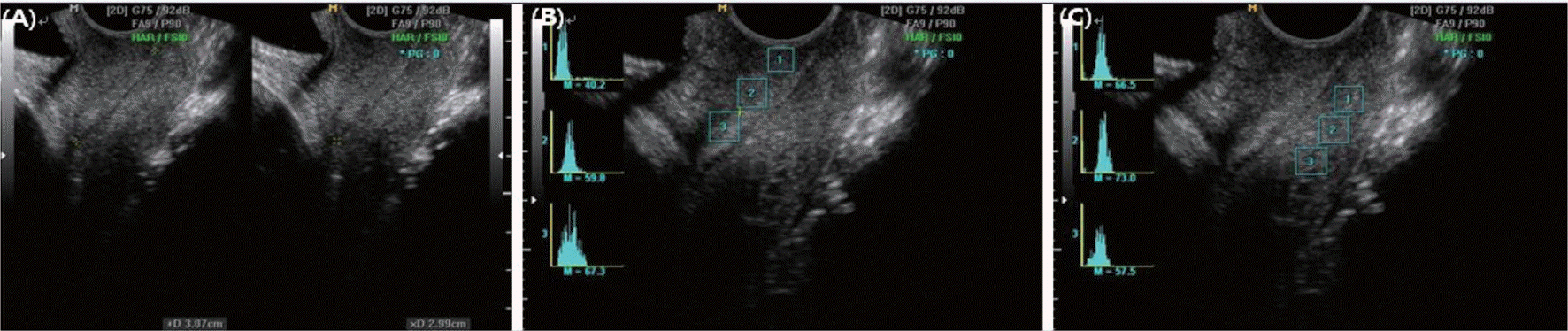 | Fig. 1Ultrasonographic assessment of the uterine cervix. (A) Cervix length, (B) Histogram (anterior lip), (C) Histogram (posterior lip). |
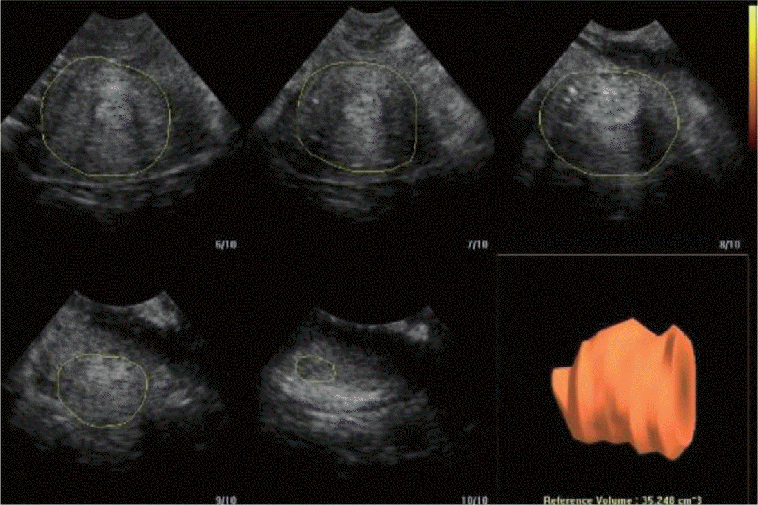 | Fig. 2Cervix volume by 3-D sonography using extended imaging virtual organ computer-aided analysis (XI VOCAL) technique. |
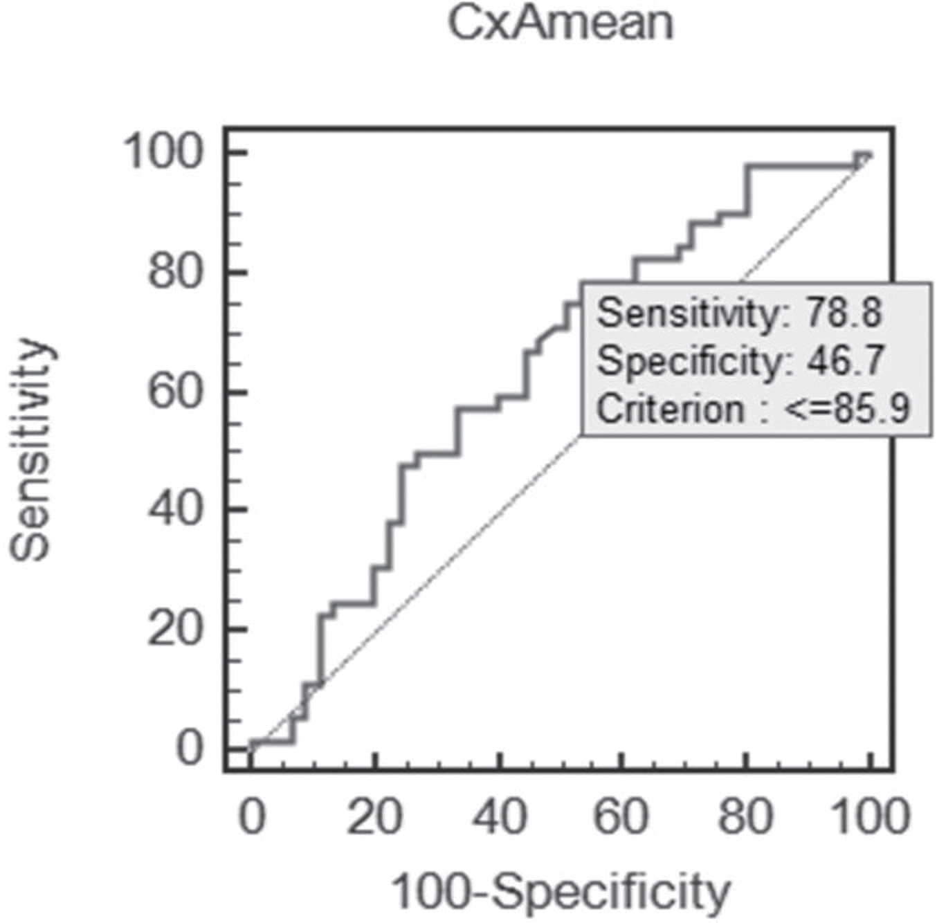 | Fig. 3Receiver operating characteristic curve for anterior lip histogram value in predicting preterm birth. The cutoff value is 85.9 (Area under the curve 0.634; Cl, 0.522-0.747; P=0.023). Abbreviation: CxAmean, Cervix anterior lip histogram mean value. |
Table 1.
Demographic and Clinical Characteristics
Table 2.
Demographic and Clinical Characteristics of Patients According to Preterm Birth
Table 3.
Factors Affecting the Probability of Preterm Birth in Patients with Preterm Labor
| Factor | Odds ratio | 95% CI |
|---|---|---|
| Bishop score | 1.250 | 0.970-1.610 |
| Cervix length | 0.189 | 0.064-0.560 |
| CxA (mean) | 0.523 | 0.168-1.627 |
| CxP (mean) | 0.752 | 0.251-2.251 |
| CxAPdiffernece | 0.503 | 0.187-1.354 |
 XML Download