

 PDF
PDF ePub
ePub Citation
 Print
Print


Abstract
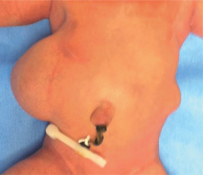
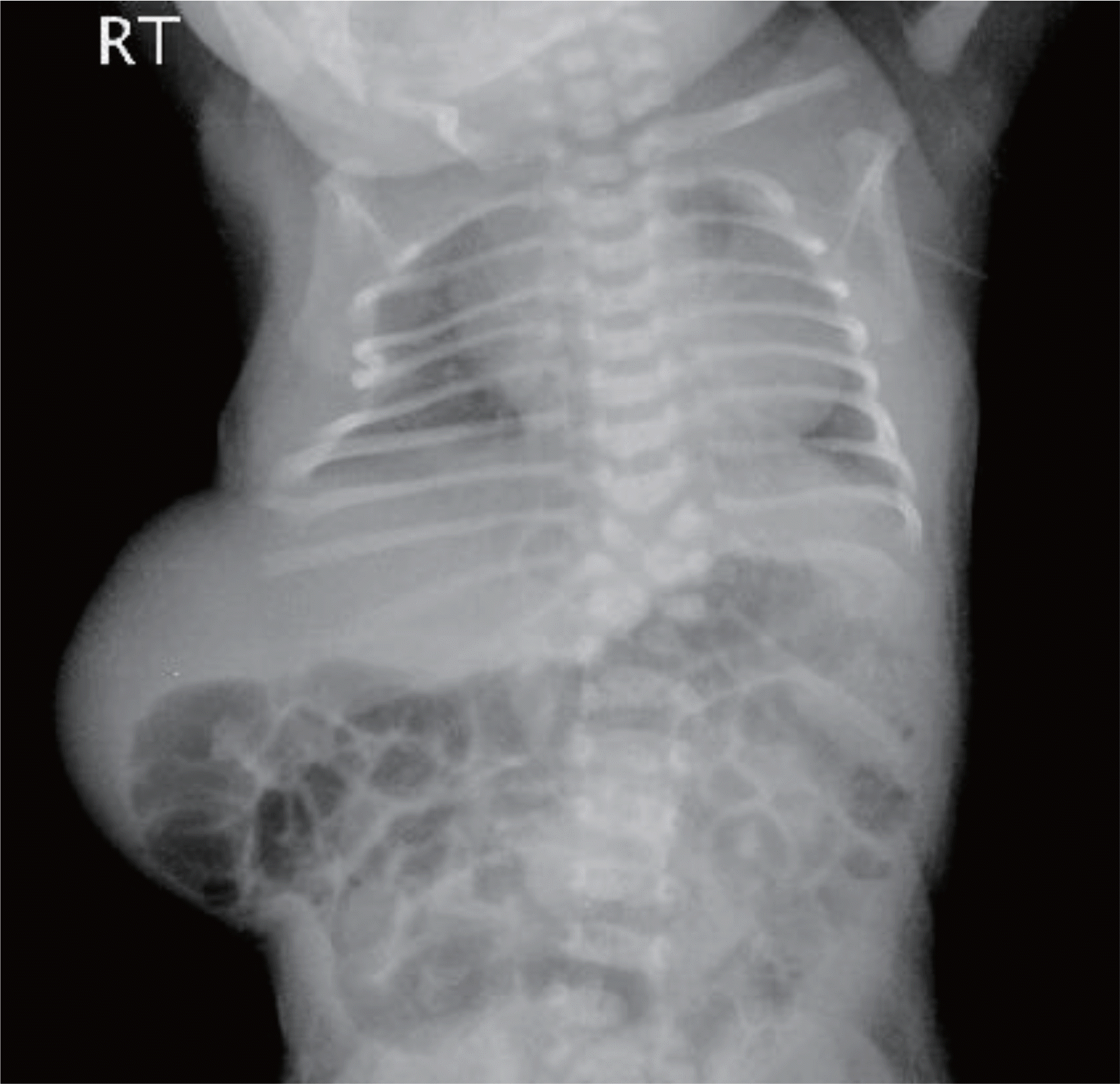
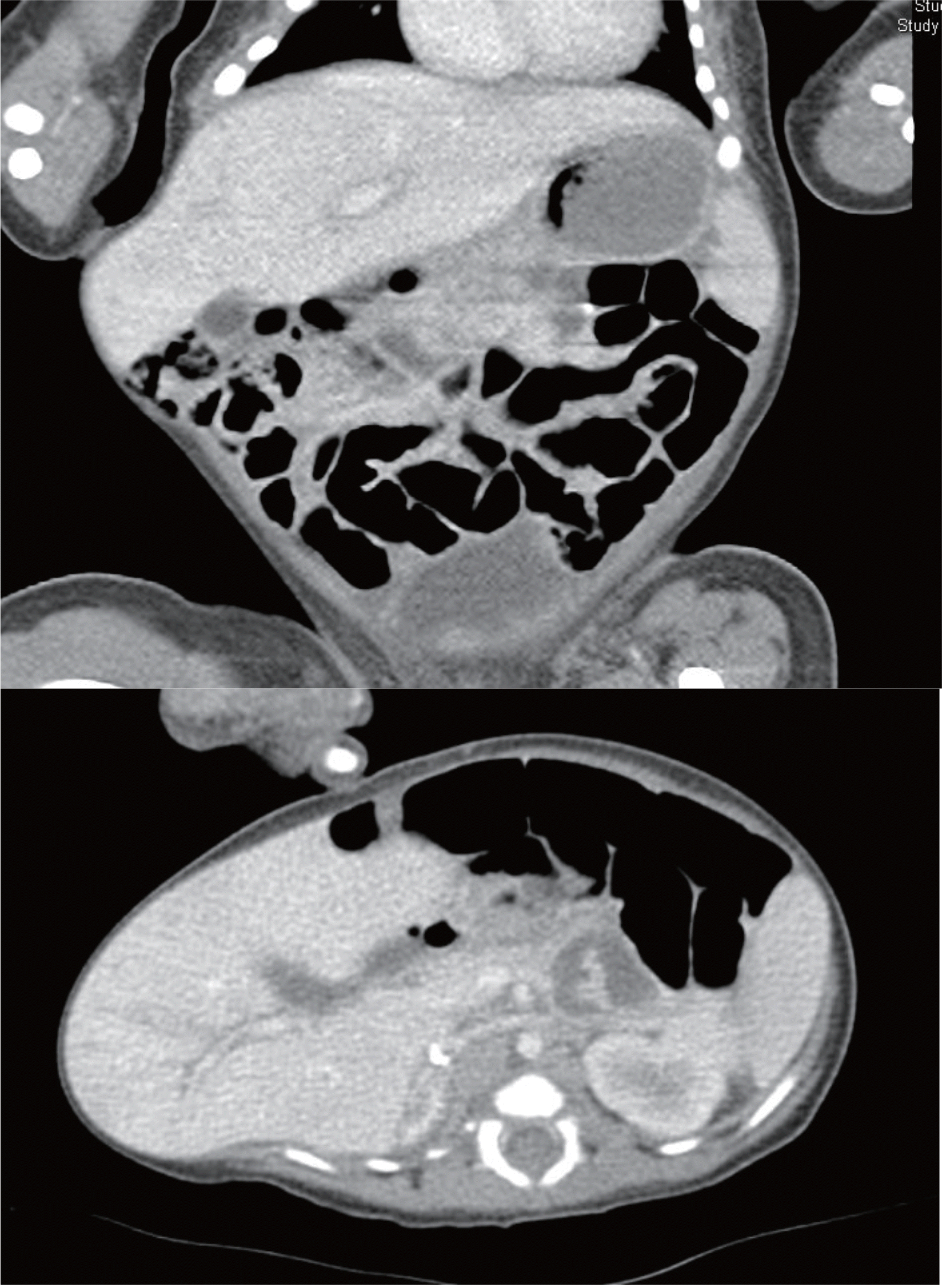
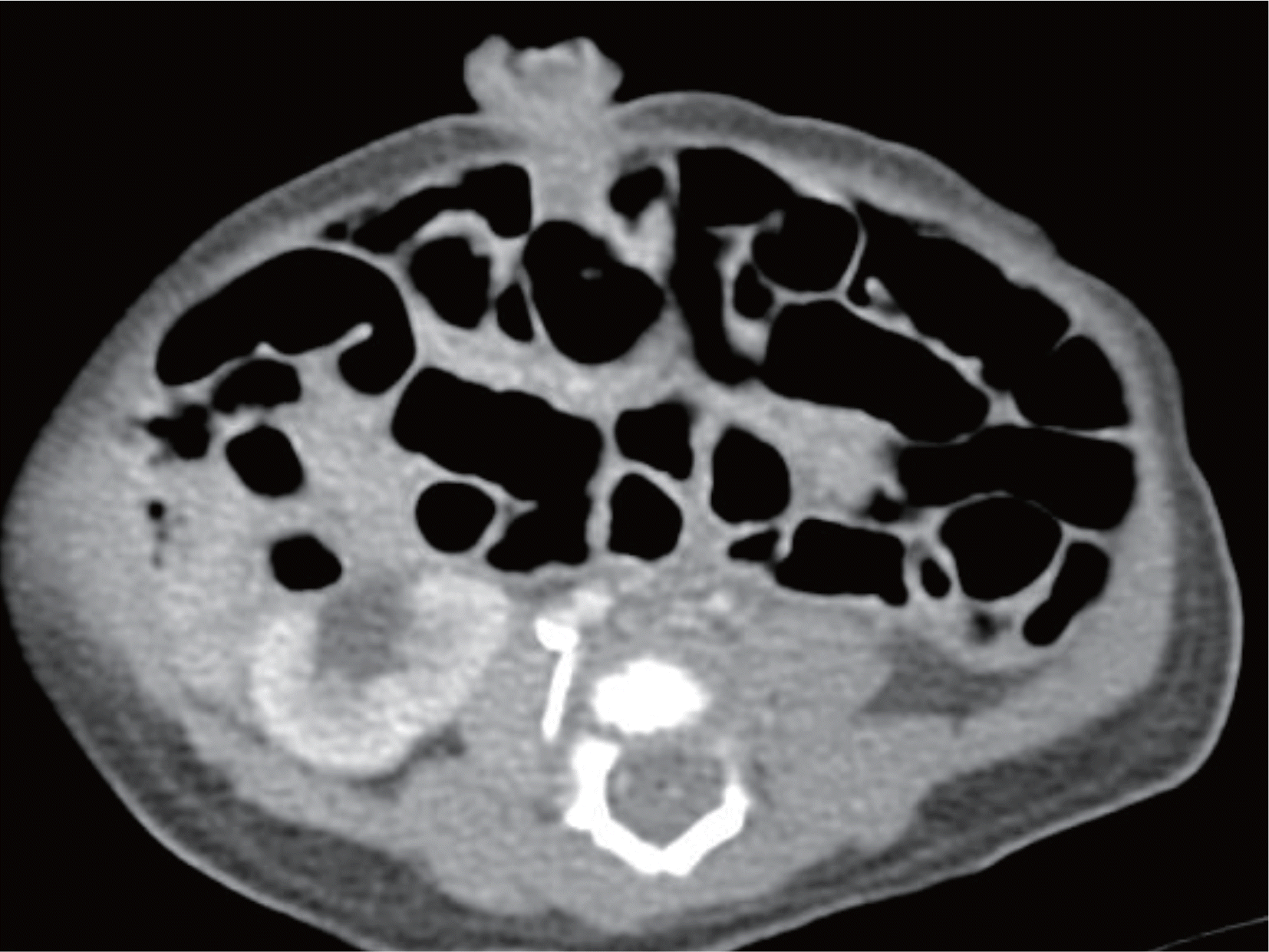
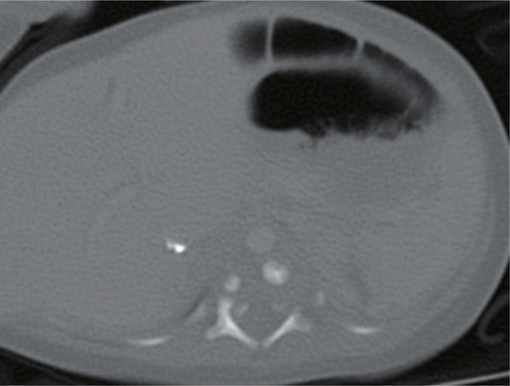
Lumbocostovertebral syndrome is a rare type of congenital lumbar hernia. Its features include lumbar hernia associated with genitourinary, vertebral, and rib anomalies. About 30 cases have been reported in the English literature, but in Korea, there has not been a case reported. We experienced a neonate with multiple costovertebral anomalies and bilateral lumbar hernia with liver and small intestine herniation diagnosed by physical examination and computed tomography. We report a case of a congenital lumbocostovertebral syndrome in neonate with literature review.
REFERENCES
1). Touloukian RJ. The lumbocostovertebral syndrome: a single somatic defect. Surgery. 1972. 71:178.
2). Carbonell AM., Kercher KW., Sigmon L., Matthews BD., Sing RF., Kneisl JS, et al. A novel technique of lumbar hernia repair using bone anchor fixation. Hernia. 2005. 9:22–6.

3). deGarangeot RJ. Colon, trainte operation Chir. 1731. 1:369–70.
4). Krishnamurthy S., Kapoor S. An incomplete form of lumbo-costovertebral syndrome in association with atrial septal defect, arthrogryposis and clubfeet. Indian J Pediatr. 2009. 76:411–3.
5). Kumar GS., Kulkarni V., Haran RP. Lumbo-costo-vertebral syndrome with posterior spinal dysraphism. Neurol India. 2005. 53:351–3.
6). Harris K., Dorn C., Bloom B. Lumbocostovertebral syn- drome with associated VACTERL anomalad: a neonatal case report. J Perinatol. 2009. 29:826–7.
7). Lafer DJ. Neuroblastoma and lumbar hernia: a causal relationship? J Pediatr Surg. 1994. 29:926–9.
8). Wakhlu A., Wakhlu AK. Congenital lumbar hernia. Pediatr Surg Int. 2000. 16:146–8.
9). Lee CM Jr., Mattheis H. Congenital lumbar hernia. Arch Dis Child. 1957. 32:42–7.
10). Skrekas G., Stafyla VK., Papalois VE. A Grynfeltt hernia: report of a case. Hernia. 2005. 9:188–91.
11). Armstrong O., Hamel A., Grignon B., NDoye JM., Hamel O., Robert R, et al. Lumbar hernia: anatomical basis and clinical aspects. Surg Radiol Anat. 2008. 30:533–7.
12). Cavallaro G., Sadighi A., Miceli M., Burza A., Carbone G., Cavallaro A. Primary lumbar hernia repair: the open approach. Eur Surg Res. 2007. 39:88–92.
13). al-Salem AH., Abu-Srair H., Qaissaruddin S. Focal nodular hyperplasia of the liver with the lumbo-costovertebral syndrome. J Pediatr Surg. 1996. 31:1282–4.
 XML Download