

 PDF
PDF ePub
ePub Citation
 Print
Print


Abstract
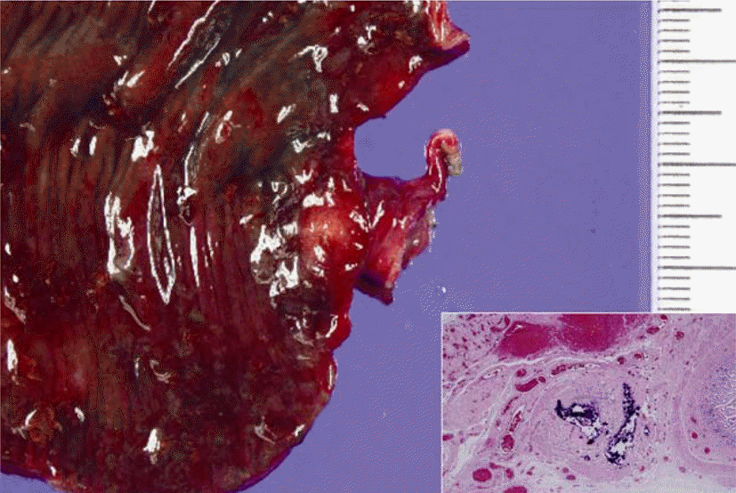
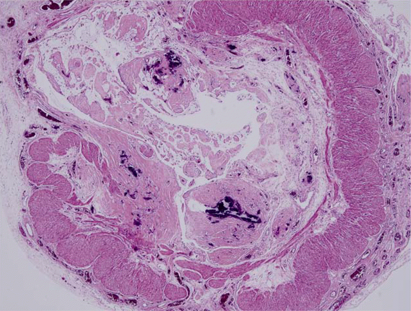
An isolated tubular intestinal loop (ITIL) means an anatomical or vascular communication with rest of the bowel loop and may provide an insight into the pathogenesis of intestinal atresia. We experienced a case of an ITIL identified in omentum of a 4-day-old neonate with type-II intestinal atresia. To our knowledge, this association has never been reported in the English literature. Omental wrapping of the incompletely resorbed ischemic bowel segment may explain this phenomenon in a case of congenital intestinal atresia.
REFERENCES
1). Louw JH., Barnard CN. Congenital intestinal atresia; observations on its origin. Lancet. 1955. 269:1065–7.

2). Nixon HH., Tawes R. Etiology and treatment of small intestinal atresia: analysis of a series of 127 jejunoileal atresias and comparison with 62 duodenal atresias. Surgery. 1971. 69:41–51.
3). Dalla Vecchia LK., Grosfeld JL., West KW., Rescorla FJ., Scherer LR., Engum SA. Intestinal atresia and stenosis: a 25-year experience with 277 cases. Arch Surg. 1998. 133:490–6. discussion 496-7.
4). Frischer J., Azizkhan R. Jejunoileal Atresia and Stenosis, in Pediatric Surgery. 7th ed.ed. Edited by Coran AG, Philadelphia, PA: Elsevier/Saunders;2012. p. p. 1059–71.
5). Abrams JS. Experimental intestinal atresia. Surgery. 1968. 64:185–91.
6). Dickinson SJ. Origin of intestinal atresia of newborn. JAMA. 1964. 190:119–21.
7). Shoshany G., Cohen E., Mordohovich D., Hayari L., Har-Shai Y., Bar-Maor JA. Creation of the isolated bowel segment in animals by omentoenteropexy. J Pediatr Surg. 1994. 29:1344–7.
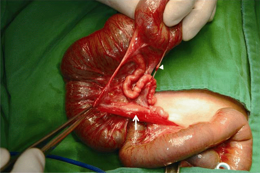
Fig. 1
Operative findings of intestinal atresia showing type I atresia and a tubular structure (indicated by arrow) embedded in omentum, and a cord-like band (indicated by arrowhead).
 XML Download